

 July 12th, 2015
  Riffin
 Ctenophores are traditionally regarded as “lower” metazoans, sharing with cnidarians a diploblastic grade of organization. Unlike cnidarians, where skeletonization (biomineralization and sclerotization) evolved repeatedly among ecologically important taxa (for example, scleractinians and octocorals), living ctenophores are characteristically soft-bodied animals. We report six sclerotized and armored ctenophores from the early Cambrian period. They have diagnostic ctenophore features (for example, an octamerous symmetry, oral-aboral axis, aboral sense organ, and octaradially arranged ctene rows). Unlike most modern counterparts, however, they lack tentacles, have a sclerotized framework, and have eight pairs of ctene rows. They are resolved as a monophyletic group (Scleroctenophora new class) within the ctenophores. This clade reveals a cryptic history and sheds new light on the early evolution of this basal animal phylum. Skeletonization also occurs in some other Cambrian animal groups whose extant members are exclusively soft-bodied, suggesting the ecological importance of skeletonization in the Cambrian explosion.
Science Advances 10 Jul 2015:Vol. 1, no. 6, e1500092,DOI: 10.1126/sciadv.1500092
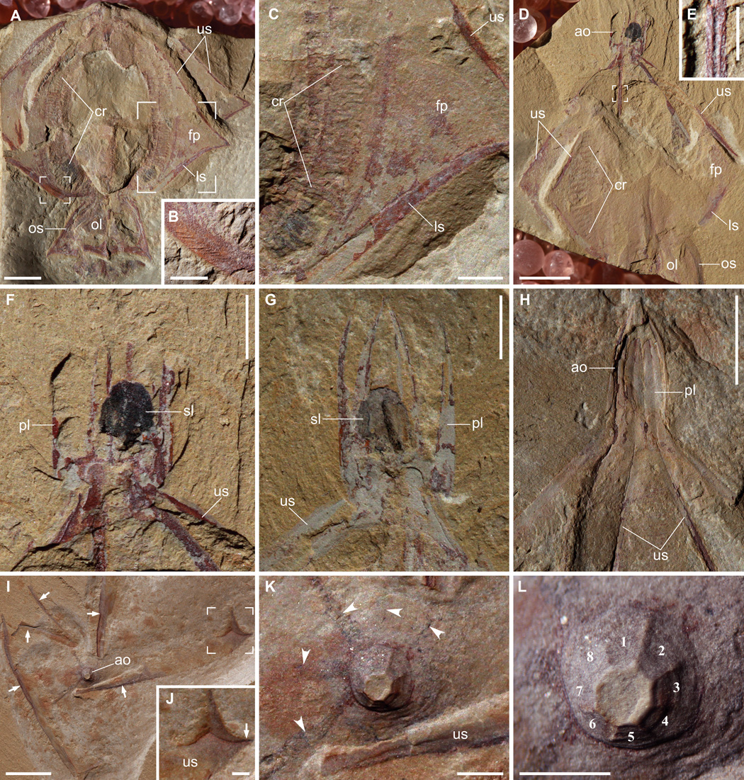
Gemmactena actinala gen. et sp. nov. (A) Holotype (ELEL-SJ100756A) showing radiating flap-like structures outlined by spokes, comb rows, and oral lappets. Apical organ not preserved. (B) Close-up of small focus area in (A) showing fine striae on spoke surface. (C) Close-up of large focus area in (A) showing remains of comb rows, as well as upper and lower spokes that frame a flap. (D) Paratype (ELEL-SJ081292A). (E) Close-up of focus area in (D) showing rigidity of a spoke with a medial groove (internal view). (F) Close-up of aboral region in (D) showing an ellipsoidal statolith preserved as dark remains surrounded by apical plates. (G) Counterpart of (F) showing complete apical plates detached from each other; remains of organic carbon are partially preserved as a dark band on the statolith. (H) Paratype (ELEL-SJ081366A) showing pointed dome-like apical organ walled by rigid plates (each with a medial groove continuous with a spoke). (I) Aborally compacted specimen ELEL-SJ120375A showing an apical organ and partially dislocated upper spokes (arrows). The apex of the apical organ was truncated and retained in counterpart during splitting. (J) Close-up of focus area in (I) showing distal end of upper spoke, with the kink marked by arrow. (K) Close-up of apical organ in (I); dark, equally spaced bands (arrowheads) radiating from the base of apical organ may represent remains of underlying meridional canals. (L) Close-up of apical organ showing considerable positive relief, presumably due to its rigidity imparted by apical plates (numbered). ao, apical organ; cr, comb row; fp, flap-like structure; ls, lower spoke; ol, oral lappet; os, oral skirt; pl, apical plate; sl, statolith; us, upper spoke. Scale bars, 5 mm (A, D, H, and I); 2 mm (C, F, and G); 1 mm (B, E, and J to L).
 Posted in
  Tags:
Tags: 