

 May 7th, 2016
  Riffin
 Highlighting and identifying fossilized structures can be difficult whether it is bone, soft tissue such as skin, muscle and internal organs, or integument such as scales and feathers. Historically, multiple methods have been used to highlight structures for photography, including cross-lighting, polarized light , camera filters, and ultraviolet (UV) light . Cross-lighting can highlight structures that are difficult to see in direct light. Polarized light can help to enhance image contrast. UV light is capable of causing minerals (e.g. bone [hydroxyapatite]) to fluoresce, and can even highlight soft tissue to some extent . This paper describes a next-generation method of fluorescing minerals using specific wavelengths of light produced by a laser and corresponding imaging through the use of laser-blocking longpass camera filters . This method is herein named Laser-stimulated fluorescence (LSF).
Feather under reflected and matrix fluoresced illumination.
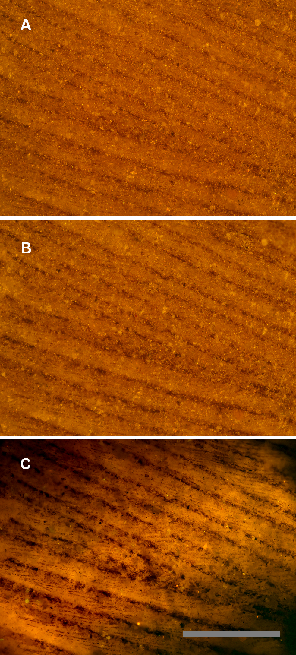
Green River Formation feather using identical images under different lighting conditions. A, Reflected light microscopy, only barbs are visible. B, Polarized light, some traces of barbules. C, Laser-stimulated fluorescence of matrix behind the carbon film backlights the feather and renders barbules visible across the entire field of view. Scale bar 0.5 mm.
For many decades UV light has been used at night to find and collect fluorescent mineral specimens, which are prized for their wide variation in color . The field of biology has made tremendous scientific advances through the use of laser-induced fluorescence mostly through the widespread use of confocal laser microscopes . In paleontology, UV light has seen increasing use in recent years where the resulting fluorescence can often reveal structures and patterns not seen under white light. The typical UV light source consists of commonly available standard fluorescent lamps with low wattage and a wavelength of 364 nanometers (nm) . Greater amounts of UV flux on the specimen will cause fluorescent minerals to become more conspicuous, allowing for easier photographic documentation, sometimes with the aid of UV filters (e.g. Hoya brand) . The limited variety of detectable fluorescence in fossils has been a primary limitation in the past using standard UV bulbs .
Feather structure comparison using white light, polarized and laser illumination.
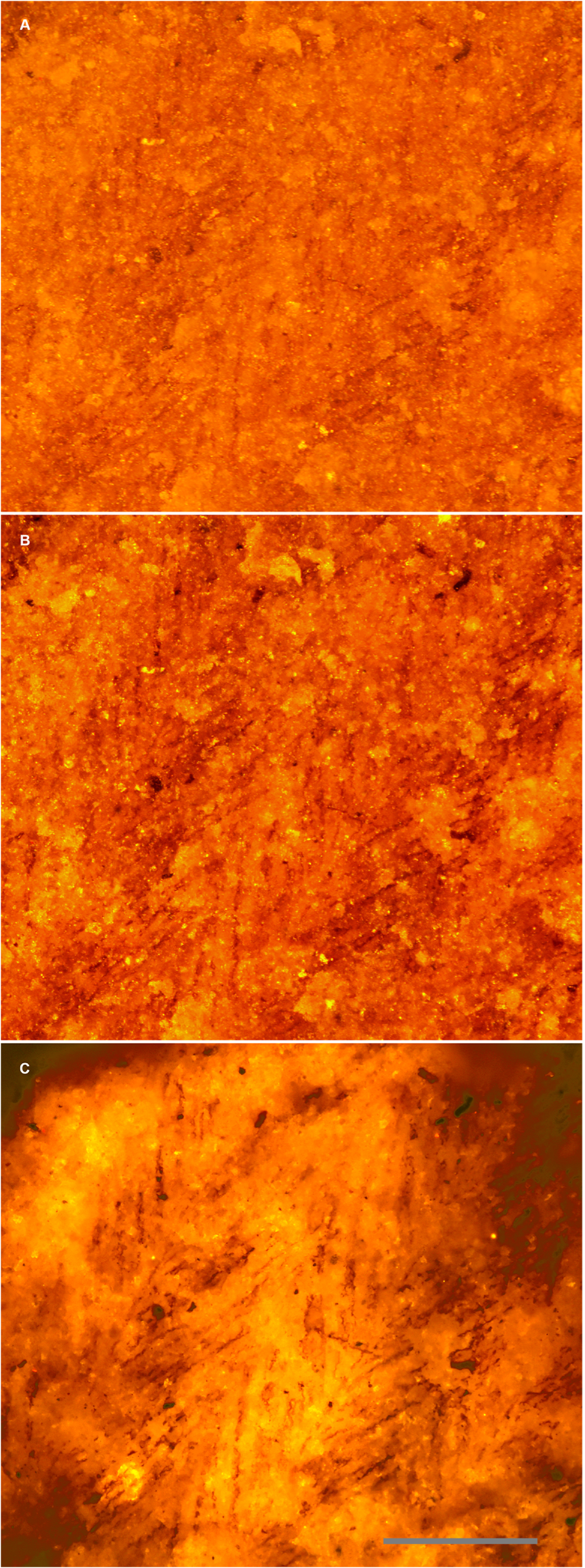
A second Green River Formation feather specimen under: A, white light, B, polarized light, and C, laser illumination. Scale bar 0.2 mm.
The technique presented here utilizes laser illumination to stimulate fluorescence which offers an order of magnitude improvement in the signal-to-noise ratio over standard UV light. The irradiance of a 20 watt UV fluorescent lamp is about 510 milliwatts per square centimeter (mWcm-2) at a distance of 20 centimeters from the target , but the irradiance of a ½ watt laser is on the order of 4000–8000 mWcm-2 . This results in detectable fluorescence of many hard-to-fluoresce mineral types which typically remain dark under standard UV. This advantage can be leveraged when other factors are accounted for. For instance, matching the correct laser line with one of the specimen’s absorption bands provides more effective excitation of the fluorescence in a sample. Furthermore, using the right optical filter that matches one of the fluorescence bands of the specimen would improve contrast in the fluorescence image.
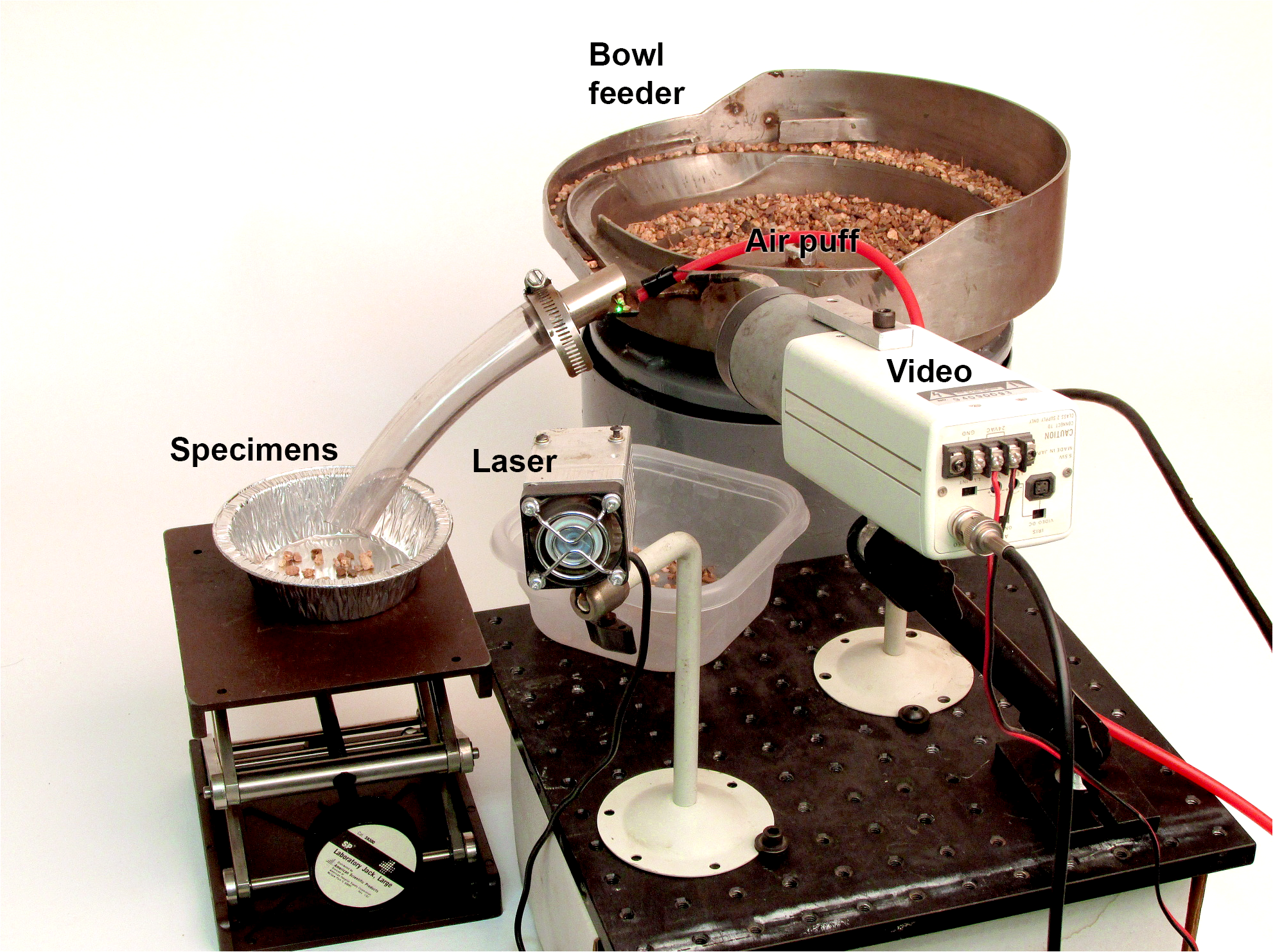
Automated fossil sorter.
Proof-of-concept prototype automated micro-fossil picker. The feeder bowl guides a stream of matrix under the laser while a video camera detects ‘blobs’ of a certain size and brightness. Fluorescing fossils are guided down a tube into a tray by a puff of compressed air
Each color of laser emits a different wavelength of light, which will excite fossils and matrix from different rock units in different ways, as the case histories that follow will indicate. Again, LSF techniques depend on the wavelength of light used, the filter used, and the inherent fluorescent properties of the rocks under study. The exact methodology used, therefore, is going to vary depending on these properties.
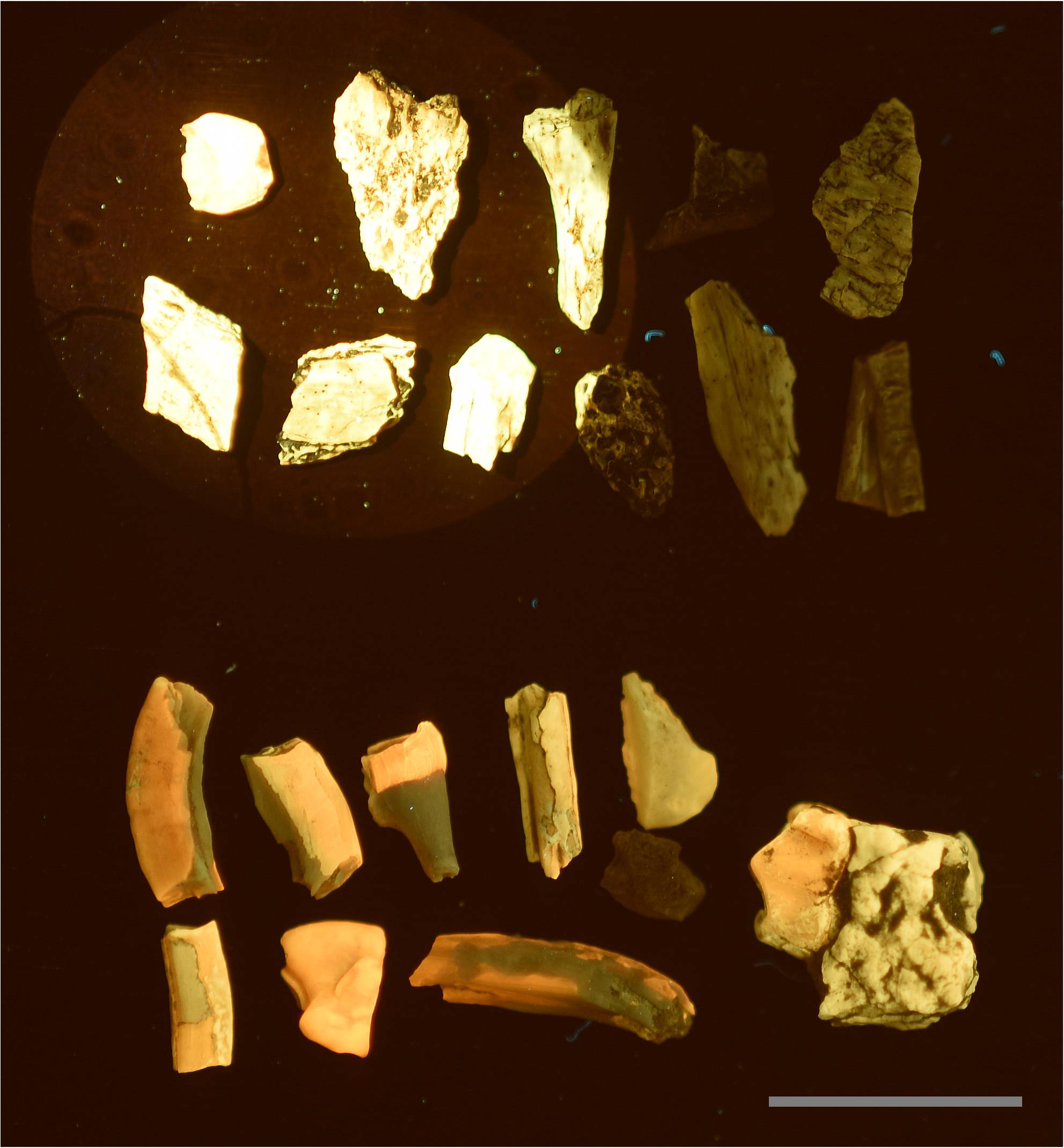
Fluorescent black light vs.blue laser. A direct comparison between a 15 watt fluorescent UV light illuminating all the fossils at a distance of 7cm, and a 447nm 500mw laser stimulating the specimens in the upper left corner. A, Specimens from the Lance Formation exhibit very low reactivity under fluorescent UVA bulbs. B, Specimens from the White River Formation typically fluoresce very well. This demonstrates that the intensity of laser stimulation can influence low reactivity specimens to fluorescence several orders of magnitude better than specimens known to fluoresce well under UV bulbs. Scale bar 1 cm
Laser-induced fluorescence imaging performed through confocal laser-scanning microscopy (CLSM) has been used in micropaleontology to study the morphology and cellular anatomy of fossils in situ, at micron-scale resolution, and even in three-dimensions . LSF is a simplified and more accessible version of CLSM that uses simpler laser beam scanning and data acquisition systems, and lacks a confocal hole. However, the LSF technique provides its own unique advantages in studying macroscopic paleontological specimens including the compactness and low cost of its setup, its fast data acquisition rate and its high sensitivity compared to UV-stimulated fluorescence. The purpose of this paper is to describe the laser-stimulated fluorescence (LSF) imaging technique and to formalize its use in paleontology in the hope that new and more efficient modes of discovery will be possible.
Citation:Kaye TG, Falk AR, Pittman M, Sereno PC, Martin LD, Burnham DA, et al. (2015) Laser-Stimulated Fluorescence in Paleontology. PLoS ONE 10(5): e0125923. doi:10.1371/journal.pone.0125923
Key: WFS,World Fossil Society,Riffin T Sajeev,Russel T Sajeev
 Posted in
  Tags:
Tags: 