

 March 9th, 2018
  Riffin
 @WFS,World Fossil Society,Riffin T Sajeev,Russel T Sajeev
Imagine that you’re a voracious carnivore who sinks its teeth into the tail of a small reptile and anticipates a delicious lunch, when, in a flash, the reptile is gone and you are left holding a wiggling tail between your jaws.
A new study by the University of Toronto Mississauga research team led by Professor Robert Reisz and PhD student Aaron LeBlanc, published March 5 in the open source journal, Scientific Reports, shows how a group of small reptiles who lived 289 million years ago could detach their tails to escape the grasp of their would-be predators — the oldest known example of such behaviour. The reptiles, called Captorhinus, weighed less than 2 kilograms and were smaller than the predators of the time. They were abundant in terrestrial communities during the Early Permian period and are distant relatives of all the reptiles today.

Fracture planes in captorhinid caudal vertebrae. (a) Artist’s reconstruction of the Permian reptile Captorhinus with an autotomous tail (inset showing anterior caudal vertebrae with fracture planes). (b) Image and (c) SEM image of an isolated anterior caudal vertebra (ROM 73769) with a fracture plane passing through the centrum (black arrow). (d) Ventral view of an anterior, rib-bearing caudal vertebra (ROM 77410) showing the absence of any fracture plane. (e) Ventral view of a caudal vertebra bearing a fracture plane (black arrows) (ROM 73771) (f) thin-section through the sagittal plane of a caudal vertebra (ROM 73773) with a fracture plane (black arrow) passing through the ventral portion of the centrum. (g) Close-up of fracture plane (black arrows) in (f) passing into the notochordal canal. Abbreviations: cb, cortical bone; cct, calcified cartilage; ce, centrum; nc, neural canal; ns, neural spine; ntc, notochordal canal. Reconstruction by Danielle Dufault. Anterior is to the left in all of the images.
As small omnivores and herbivores, Captorhinus and its relatives had to scrounge for food while avoiding being preyed upon by large meat-eating amphibians and ancient relatives of mammals. “One of the ways captorhinids could do this,” says first author LeBlanc, “was by having breakable tail vertebrae.” Like many present-day lizard species, such as skinks, that can detach their tails to escape or distract a predator, the middle of many tail vertebrae had cracks in them.
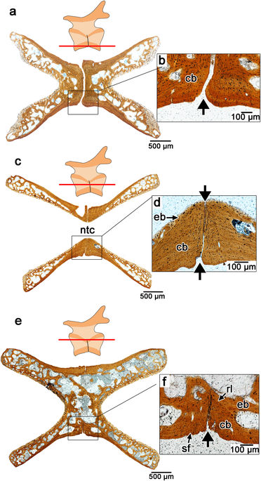
Serial transverse sections through the centra of fracture plane-bearing caudal vertebrae in captorhinids. (a) Transverse section taken near the base of the centrum (ROM 73771); (b) Closeup of the fracture plane running across the ventral surface of the centrum in (a). (c) Transverse section taken at the level of the notochordal canal (ROM 73774). (d) Closeup of the fracture plane in c passing through the centrum into the notochordal canal. (e) Transverse section taken along the roof of the centrum (below the neural arch) (ROM 73774). (f) Closeup of fracture plane in (g) passing through the outer cortical bone, but not the more internal endosteal/endochondral bone. Abbreviations: cb, cortical bone; eb, endochondral bone; rl, reversal line; sf, Sharpey’s fibers. Anterior is to the left in all of the images.
It is likely that these cracks acted like the perforated lines between two paper towel sheets, allowing vertebrae to break in half along planes of weakness. “If a predator grabbed hold of one of these reptiles, the vertebra would break at the crack and the tail would drop off, allowing the captorhinid to escape relatively unharmed,” says Reisz, a Distinguished Professor of Biology at the University of Toronto Mississauga.
The authors note that being the only reptiles with such an escape strategy may have been a key to their success, because they were the most common reptiles of their time, and by the end of the Permian period 251 million years ago, captorhinids had dispersed across the ancient supercontinent of Pangaea. This trait disappeared from the fossil record when Captorhinus died out; it re-evolved in lizards only 70 million years ago.
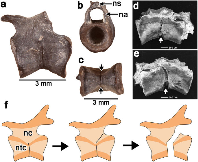
Hypothesized path of autotomy break in the caudal centra of captorhinids. (a–c) Lateral, anterior, and ventral views of a fracture plane-bearing caudal centrum, showing the extend of the fracture plane, which does not pass through the neural arch or spine (ROM 73774). (d,e) SEM images of a post mortem break in a captorhinid caudal vertebra (ROM 77409) along its fracture plane. The break follows the plane of weakness formed by the autotomy plane (white arrow) and is found on the left (d) and right (e) sides, suggesting that this was the plane of weakness in life. (f) Reconstruction of caudal autotomy in a caudal vertebra. During autotomy, the fracture follows the path of least resistance through the dorsal half of the caudal centrum and the posterior base of the neural arch, bypassing the neural spine. Abbreviation: na, neural arch; nc, neural canal; ns, neural spine; ntc, notochordal canal. Anterior is to the left in all of the images.
They were able to examine more than 70 tail vertebrae — both juveniles and adults — and partial tail skeletons with splits that ran through their vertebrae. They compared these skeletons to those of other reptilian relatives of captorhinids, but it appears that this ability is restricted to this family of reptiles in the Permian period.
Using various paleontological and histological techniques, the authors discovered that the cracks were features that formed naturally as the vertebrae were developing. Interestingly, the research team found that young captorhinids had well-formed cracks, while those in some adults tended to fuse up. This makes sense, since predation is much greater on young individuals and they need this ability to defend themselves.
This study was possible thanks to the treasure trove of fossils available at the cave deposits near Richards Spur, Oklahoma.
- A. R. H. LeBlanc, M. J. MacDougall, Y. Haridy, D. Scott, R. R. Reisz. Caudal autotomy as anti-predatory behaviour in Palaeozoic reptiles. Scientific Reports, 2018; 8 (1) DOI: 10.1038/s41598-018-21526-3
@WFS,World Fossil Society,Riffin T Sajeev,Russel T Sajeev
 Posted in
  Tags:
Tags: 