WFS News: Tharosaurus indicus, an oldest long-necked dinosaur unearthed in India

@WFS,World Fossil Society,Athira,Riffin T Sajeev,Russel T Sajeev

Fossils of the oldest long-necked dinosaur that inhabited Earth about 167 million years ago have been unearthed in India that reveal the country was a “major centre of dinosaur evolution”, experts said.

The discovery unearths some of the oldest plant-eating giant reptiles to have roamed the planet.

The dinosaur, named Tharosaurus indicus after India’s Thar desert, walked the planet during the Middle Jurassic period, according to a new study published in the journal Scientific Reports.

Researchers, including those from the Indian Institute of Technology (IIT) Roorkee and the Geological Survey of India dug up the fossil at a site near the city of Jaisalmer in the western state of Rajasthan – a region that was part of a shoreline along the prehistoric Tethys Ocean.

The new dinosaur was a member of the group known as dicraeosaurids that had long necks that fed on vegetation, according to the scientists.

It is also the first of this group of dinosaurs to have ever been found in India – and the oldest in the world.

“Prof. Sunil Bajpai and Debajit Dutta of IIT-Roorkee and Geological Survey of India have discovered the oldest fossil remains of a long-necked, plant-eating dicraeosaurid dinosaur in Jaisalmer, suggesting that India was a major centre of dinosaur evolution,” said IIT Roorkee in a post on X, formerly Twitter.

The discovery provides fresh insights into the diversity of sauropod dinosaurs in the prehistoric landmass, to which the Indian subcontinent was also a part at the time.

“Palaeobiogeographic considerations of Tharosaurus, seen in conjunction with the other Indian Jurassic sauropods, suggest that the new Indian taxon is a relic of a lineage that originated in India and underwent rapid dispersal across the rest of Pangaea,” scientists wrote in the study.

Other dinosaurs belonging to this group from the Middle Jurassic–Early Cretaceous periods have mostly been unearthed from Africa and South America as well as from sites in the US and China.

Researchers said the larger group of these dinosaurs, known as diplodocoids, all had long bodies and necks with spikes on the backs of their necks.

T indicus, they said, also slightly differed from others in its group with long depression on the side of its neck bones as well as neural spines, indicating it likely had unique spikes.

Scientists speculate the diplodocoid dinosaur group likely spread from, or may have originated in India, but added that this theory “still needs to be reconciled” with by comparing another Asian dinosaur group, the Lingwulong, from the Middle Jurassic period.

The new discovery, researchers said, also emphasises the need for increased sampling of older fossil sites in India in search of such ancestral dinosaur groups.
Source: Article by Vishwam Sankaran , www.independent.co.uk
@WFS,World Fossil Society,Athira,Riffin T Sajeev,Russel T Sajeev

WFS News: Historical RNA expression profiles from the extinct Tasmanian tiger.

@WFS,World Fossil Society,Athira,Riffin T Sajeev,Russel T Sajeev

A new study shows the isolation and sequencing of more than a century-old RNA molecules from a Tasmanian tiger specimen preserved at room temperature in a museum collection. This resulted in the reconstruction of skin and skeletal muscle transcriptomes from an extinct species for the first time. The researchers note that their findings have relevant implications for international efforts to resurrect extinct species, including both the Tasmanian tiger and the woolly mammoth, as well as for studying pandemic RNA viruses.

The Tasmanian tiger, also known as the thylacine, was a remarkable apex carnivorous marsupial that was once distributed all across the Australian continent and the island of Tasmania. This extraordinary species found its final demise after European colonization, when it was declared as an agricultural pest and a bounty of £1 per each full-grown animal killed was set by 1888. The last known living Tasmanian tiger died in captivity in 1936 at the Beaumaris Zoo in Hobart, Tasmania.

Recent efforts in de-extinction have focused on the Tasmanian tiger, as its natural habitat in Tasmania is still mostly preserved, and its reintroduction could help recovering past ecosystem equilibriums lost after its final disappearance. However, reconstructing a functional living Tasmanian tiger not only requires a comprehensive knowledge of its genome (DNA) but also of tissue-specific gene expression dynamics and how gene regulation worked, which are only attainable by studying its transcriptome (RNA).

“Resurrecting the Tasmanian tiger or the woolly mammoth is not a trivial task, and will require a deep knowledge of both the genome and transcriptome regulation of such renowned species, something that only now is starting to be revealed,” says Emilio Mármol, the lead author of a study recently published in the Genome Research journal by researchers at SciLifeLab in collaboration with the Centre for Palaeogenetics, a joint venture between the Swedish Museum of Natural History and Stockholm University.

RNA molecules recovered from the Tasmanian tiger

The researchers behind this study have sequenced, for the first time, the transcriptome of the skin and skeletal muscle tissues from a 130-year-old desiccated Tasmanian tiger specimen preserved at room temperature in the Swedish Museum of Natural History in Stockholm. This led to the identification of tissue-specific gene expression signatures that resemble those from living extant marsupial and placental mammals.

The recovered transcriptomes were of such good quality that it was possible to identify muscle- and skin-specific protein coding RNAs, and led to the annotation of missing ribosomal RNA and microRNA genes, the later following MirGeneDB recommendations.

“This is the first time that we have had a glimpse into the existence of thylacine-specific regulatory genes, such as microRNAs, that got extinct more than one century ago,” says Marc R. Friedländer, Associate Professor at the Department of Molecular Biosciences, The Wenner-Gren Institute at Stockholm University and SciLifeLab.

This pioneering study opens up new exciting opportunities and implications for exploring the vast collections of specimens and tissues stored at museums across the globe, where RNA molecules might await to be uncovered and sequenced.

“In the future, we may be able to recover RNA not only from extinct animals, but also RNA virus genomes such as SARS-CoV2 and their evolutionary precursors from the skins of bats and other host organisms held in museum collections,” says Love Dalén, Professor of evolutionary genomics at Stockholm University and the Centre for Palaeogenetics.

The authors of the study say they are excited for future holistic research developments integrating both genomics and transcriptomics towards a new era in palaeogenetics beyond DNA.

Journal Reference:

  1. Emilio Mármol-Sánchez, Bastian Fromm, Nikolay Oskolkov, Zoé Pochon, Panagiotis Kalogeropoulos, Eli Eriksson, Inna Biryukova, Vaishnovi Sekar, Erik Ersmark, Björn Andersson, Love Dalén, Marc R. Friedländer. Historical RNA expression profiles from the extinct Tasmanian tigerGenome Research, 2023; DOI: 10.1101/gr.277663.123
Stockholm University. “RNA for the first time recovered from an extinct species.” ScienceDaily. ScienceDaily, 19 September 2023. <www.sciencedaily.com/releases/2023/09/230919153758.htm>.
@WFS,World Fossil Society,Athira,Riffin T Sajeev,Russel T Sajeev

WFS News:Plate tectonic cross-roads: Reconstructing the Panthalassa-Neotethys Junction Region from Philippine Sea Plate and Australasian oceans and orogens.

@WFS,World Fossil Society, Athira, Riffin T Sajeev, Russel T Sajeev
Utrecht University geologist Suzanna van de Lagemaat has reconstructed a massive and previously unknown tectonic plate that was once one-quarter the size of the Pacific Ocean. Her colleagues in Utrecht had predicted its existence over 10 years ago based on fragments of old tectonic plates found deep in the Earth’s mantle. Van de Lagemaat reconstructed lost plates through field research and detailed investigations of the mountain belts of Japan, Borneo, the Philippines, New Guinea, and New Zealand. To her surprise, she found that oceanic remnants on northern Borneo must have belonged to the long-suspected plate, which scientists have named Pontus. She has now reconstructed the entire plate in its full glory. Suzanna van de Lagemaat will defend her dissertation on this plate tectonics puzzle at Utrecht University on Friday, October 13.

Understanding the movements of the tectonic plates that make up the earth’s rigid outer shell is essential to understand the planet’s geological history. The movements of these plates strongly influenced how the planet’s paleogeography and climate have changed over time, and even where to find rare metals. But large oceanic plates from the geological past have since disappeared into the earth’s mantle by means of subduction. They have left behind only fragments of rock hidden in mountain belts. Van de Lagemaat studied the planet’s most complicated plate tectonic region: the area around the Philippines. “The Philippines is located at a complex junction of different plate systems. The region almost entirely consists of oceanic crust, but some pieces are raised above sea level, and show rocks of very different ages.”

Reconstruction

Using geological data, Van de Lagemaat first reconstructed the movements of the current plates in the region between Japan and New Zealand. That revealed how large the area was of plates that must have disappeared in the current western Pacific region. “We also conducted field work on northern Borneo, where we found the most important piece of the puzzle. We thought we were dealing with relicts of a lost plate that we already knew about. But our magnetic lab research on those rocks indicated that our finds were originally from much farther north, and had to be remnants of a different, previously unknown plate.” But the important realisation was yet to come. “11 years ago, we thought that the remnants of Pontus might lie in northern Japan, but we’d since refuted that theory,” explains Douwe van Hinsbergen, Van de Lagemaat’s PhD supervisor. “It was only after Suzanna had systematically reconstructed half of the ‘Ring of Fire’ mountain belts from Japan, through New Guinea, to New Zealand that the proposed Pontus plate revealed itself, and it included the rocks we studied on Borneo.”

Relics

The relics of Pontus are not only located on northern Borneo, but also on Palawan, an island in the Western Philippines, and in the South China Sea. Van de Lagemaat’s research also showed that a single coherent plate tectonic system stretched from southern Japan to New Zealand, and it must have existed for at least 150 million years. That is also a new discovery in the field.

Waves

The previous predictions of the existence of Pontus were made possible because a subducted plate leaves behind traces when it ‘sinks’ into the earth’s mantle: zones in the mantle with anomalous temperatures or compositions. These anomalies can be observed when seismographs pick up signals from earthquakes. Earthquakes send waves through Earth’s interior, and when they travel through an anomaly, such as a fragment from an old plate, the anomaly produces a disruption of the signal. Geologists can trace these disruptions to the existence of phenomena in the mantle, such as fragments of tectonic plates. That allows them to look 300 million years into the past; older plate fragments have ‘dissolved’ at the boundary between the mantle and the core. The study from 11 years ago showed that a large subduction zone must have run through the western paleo-Pacific Ocean, which separated the known Pacific plates in the east from the hypothetical Pontus plate in the west. This hypothesis has now been independently demonstrated by Van de Lagemaat’s research.

Journal Reference:

  1. Suzanna H.A. van de Lagemaat, Douwe J.J. van Hinsbergen. Plate tectonic cross-roads: Reconstructing the Panthalassa-Neotethys Junction Region from Philippine Sea Plate and Australasian oceans and orogensGondwana Research, 2023; DOI: 10.1016/j.gr.2023.09.013
Utrecht University. “Plate tectonic surprise: Geologist unexpectedly finds remnants of a lost mega-plate.” ScienceDaily. ScienceDaily, 9 October 2023. <www.sciencedaily.com/releases/2023/10/231009191657.htm>.
@WFS,World Fossil Society, Athira, Riffin T Sajeev, Russel T Sajeev

WFS News:125-Million-Year-Old Dinosaur Feathers Reveal Traces of Ancient Proteins

@WFS,World Fossil Society, Athira, Riffin T Sajeev, Russel T Sajeev

Paleontologists at University College Cork (UCC) in Ireland have discovered X-ray evidence of proteins in fossil feathers that sheds new light on feather evolution.

A graphical abstract based on paper by Slater et al., 2003. Credit: Science Graphic Design

A graphical abstract based on paper by Slater et al., 2003. Credit: Science Graphic Design

Previous studies suggested that ancient feathers had a different composition to the feathers of birds today. The new research, however, reveals that the protein composition of modern-day feathers was also present in the feathers of dinosaurs and early birds, confirming that the chemistry of feathers originated much earlier than previously thought.

The research, published today in Nature Ecology and Evolution, was led by paleontologists Dr. Tiffany Slater and Prof. Maria McNamara of UCC’s School of Biological, Earth, and Environmental Science, who teamed with scientists based at Linyi University (China) and the Stanford Synchrotron Radiation Lightsource (USA).

The team analyzed 125-million-year-old feathers from the dinosaur Sinornithosaurus and the early bird Confuciusornis from China, plus a 50-million-year-old feather from the USA.

“It’s really exciting to discover new similarities between dinosaurs and birds,” Dr. Slater says. “To do this, we developed a new method to detect traces of ancient feather proteins. Using X-rays and infrared light we found that feathers from the dinosaur Sinornithosaurus contained lots of beta-proteins, just like feathers of birds today.”

To help interpret the chemical signals preserved in the fossil feathers, the team also ran experiments to help understand how feather proteins break down during the fossilization process.

“Modern bird feathers are rich in beta-proteins that help strengthen feathers for flight,” Dr. Slater says

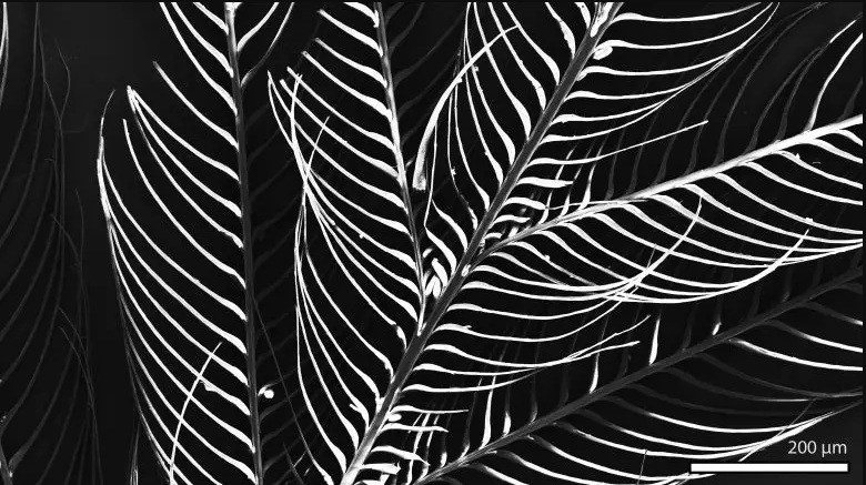
Scanning electron microscopy image of zebra finch feather. Scale bar indicates 200 µm. Credit: Dr. Tiffany Slater

Scanning electron microscopy image of zebra finch feather. Scale bar indicates 200 µm. Credit: Dr. Tiffany Slater

“Previous tests on dinosaur feathers, though, found mostly alpha-proteins. Our experiments can now explain this weird chemistry as the result of protein degradation during the fossilization process. So although some fossil feathers do preserve traces of the original beta-proteins, other fossil feathers are damaged and tell us a false narrative about feather evolution.”

This research helps answer a long-standing debate about whether feather proteins, and proteins in general, can preserve in deep time.

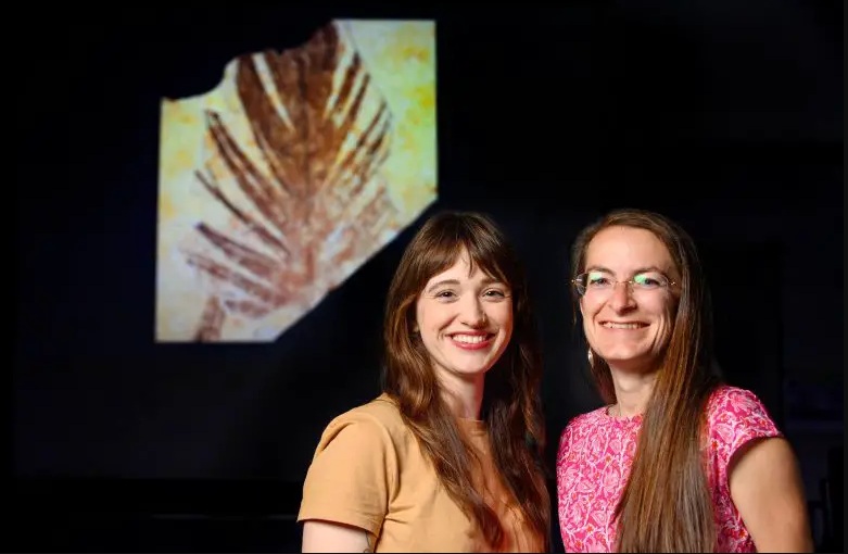
Dr. Tiffany Slater and Prof. Maria McNamara pictured in the experimental fossilization laboratory at the School of Biological, Earth and Environmental Sciences at University College Cork. Credit: Daragh Mc Sweeney/Provision

Dr. Tiffany Slater and Prof. Maria McNamara pictured in the experimental fossilization laboratory at the School of Biological, Earth and Environmental Sciences at University College Cork. Credit: Daragh Mc Sweeney/Provision

Prof. Maria McNamara, senior author on the study, said “Traces of ancient biomolecules can clearly survive for millions of years, but you can’t read the fossil record literally because even seemingly well-preserved fossil tissues have been cooked and squashed during fossilization. We’re developing new tools to understand what happens during fossilization and unlock the chemical secrets of fossils. This will give us exciting new insights into the evolution of important tissues and their biomolecules.”

Reference: “Preservation of corneous β-proteins in Mesozoic feathers” by Tiffany S. Slater, Nicholas P. Edwards, Samuel M. Webb, Fucheng Zhang, and Maria E. McNamara, 21 September 2023, Nature Ecology & Evolution.
DOI: 10.1038/s41559-023-02177-8

Source: Article  By 

@WFS,World Fossil Society, Athira, Riffin T Sajeev, Russel T Sajeev

WFS News: New Research Sheds Light on How Dinosaurs Became Giants

@WFS,World Fossil Society, Athira, Riffin T Sajeev, Russel T Sajeev

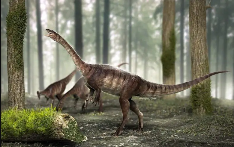
Bone cavities, known as air sacs, emerged in precursors of long-necked dinosaurs roughly 225 million years ago, as evidenced by a specimen unearthed in Rio Grande do Sul, South Brazil. The research also indicates that air sacs did not evolve as linearly as scientists believe. Credit: Márcio L. Castro

Bone cavities, known as air sacs, emerged in precursors of long-necked dinosaurs roughly 225 million years ago, as evidenced by a specimen unearthed in Rio Grande do Sul, South Brazil. The research also indicates that air sacs did not evolve as linearly as scientists believe. Credit: Márcio L. Castro

Bone cavities called air sacs emerged in the precursors of long-necked dinosaurs around 225 million years ago, according to the analysis of a specimen found in Rio Grande do Sul state, South Brazil.

The missing link has just been found, bridging the gap between the earliest dinosaurs, which varied significantly in size from mere centimeters to a maximum of 3 meters, and more recent giants that could be more than twice the length of a bus and have so much appeal to the popular imagination.

Macrocollum itaquii, which was discovered in the region of Agudo in the Rio Grande do Sul state of South Brazil and dates back 225 million years, is the most ancient dinosaur known to have structures referred to as air sacs.

These bone cavities, which persist in present-day birds, enabled dinosaurs to capture more oxygen, keep their bodies cool, and withstand the harsh conditions of their era. They also helped some become giants: Tyrannosaurus rex and Brachiosaurus, for example.

An article on the study that led to the discovery was published in the journal Anatomical Record. Two of its authors are researchers supported by FAPESP at the State University of Campinas (UNICAMP) in São Paulo state.

Skeletal reconstruction of the unaysaurid sauropodomorph Macrocollum (CAPPA/UFSM 0001b) showing vertebral elements along the spine and putative reconstruction of the air sac systems involved. (a) Pneumatic posterior cervical vertebra and a cross-section CT slice in b. (c) a pneumatized anterior dorsal vertebra with cross-section CT slice in d, and detail of the pneumatic foramen in e. (f) Detail of the pneumatic foramen in a reconstructed 3D model of the element. (g) Anterior cervical element (apneumatic). (h) Posterior dorsal vertebra shows no traces of PSP. The sacral series (i), as well as the anterior (k) and mid-caudal (j) series are apneumatic. a, g, h, j, and k are in left lateral view. c, e and f are in right lateral view. i is in dorsal view. ABD, abdominal diverticula; CER, cervical diverticula; LUN, lung; pf, pneumatic foramen. The reconstruction was made by Rodrigo T. Müller. Scale bar of the skeletal reconstruction = 500 mm; a–j = 20 mm.

Skeletal reconstruction of the unaysaurid sauropodomorph Macrocollum (CAPPA/UFSM 0001b) showing vertebral elements along the spine and putative reconstruction of the air sac systems involved. (a) Pneumatic posterior cervical vertebra and a cross-section CT slice in b. (c) a pneumatized anterior dorsal vertebra with cross-section CT slice in d, and detail of the pneumatic foramen in e. (f) Detail of the pneumatic foramen in a reconstructed 3D model of the element. (g) Anterior cervical element (apneumatic). (h) Posterior dorsal vertebra shows no traces of PSP. The sacral series (i), as well as the anterior (k) and mid-caudal (j) series are apneumatic. a, g, h, j, and k are in left lateral view. c, e and f are in right lateral view. i is in dorsal view. ABD, abdominal diverticula; CER, cervical diverticula; LUN, lung; pf, pneumatic foramen. The reconstruction was made by Rodrigo T. Müller. Scale bar of the skeletal reconstruction = 500 mm; a–j = 20 mm.

“Air sacs made their bones less dense, allowing them to grow to more than 30 meters in length,” said Tito Aureliano, first author of the article. The study was conducted as part of his PhD research at the Institute of Geosciences (IG-UNICAMP).

M. itaquii was the largest dinosaur of its time, with a length of about 3 meters. A few million years before then, the largest dinosaurs were about 1 meter long. Air sacs certainly facilitated this increase in size,” Aureliano added.

The study was a stage of the project “Taphonomic landscapes,” funded by FAPESP. Taphonomy is the study of how organisms decay and become fossilized or preserved in the paleontological record.

The principal investigator for this project was Fresia Ricardi-Branco, the penultimate author of the article and a professor at IG-UNICAMP.

“This was one of the first dinosaurs to walk the Earth, in the Triassic period,” she said. “The air sac adaptation enabled it to grow and withstand the climate in this period and later, in the Jurassic and Cretaceous. Air sacs gave dinosaurs an evolutionary advantage over other groups, such as mammals, and they were able to diversify faster.”

In a previous study, the group showed that the earliest fossils found so far did not have air sacs, taking their absence as a sign that this trait evolved at least three times independently.

M. itaquii was a biped, a sauropodomorph, and an ancestor of the giant quadrupeds with a small head, and a neck at least as long as the trunk.

Nonlinear evolution

Until air sacs were discovered in M. itaquii, these vertebral cavities were known to consist of either camerate or camellate tissue, the former referring to hollow spaces observed by microtomography, and the latter to spongy bone. According to the authors, in this case, they found “internal pneumatic chambers”, which are “neither camerate nor camellate, but a new type of tissue with an intermediate texture”. They propose to call the new structures “protocamerate”, as they “are not large enough to be considered camerae, but also present a camellate array internally”.

“The most widely held hypothesis until now was that the air sacs began as camerae and evolved into camellae. Our proposal, based on what we observed in this specimen, is that this other form existed first of all,” Aureliano said.

The vertebrae in which the air sacs were found also changed what was known about the evolution of these structures. Based on the fossils analyzed previously, other research groups proposed that air sacs first appeared in the abdominal region and did not appear in the cervical region until the early Jurassic (190 million years ago), a long time after the period in which M. itaquii was alive. Here, however, the authors found clear evidence of air sacs in the cervical and dorsal regions, with no sign of the structures in the abdominal region.

“It’s as if evolution had conducted different experiments until it arrived at the definitive system, in which air sacs run from the cervical region to the tail. It wasn’t a linear process,” Aureliano said.

Reference: “The origin of an invasive air sac system in sauropodomorph dinosaurs” by Tito Aureliano, Aline M. Ghilardi, Rodrigo T. Müller, Leonardo Kerber, Marcelo A. Fernandes, Fresia Ricardi-Branco and Mathew J. Wedel, 27 March 2023, The Anatomical Record.
DOI: 10.1002/ar.25209

Source: Report by  

@WFS,World Fossil Society, Athira, Riffin T Sajeev, Russel T Sajeev

WFS News: Long-Standing Question Answered – How Mass Extinction Paved the Way for Oysters and Clams

@WFS,World Fossil Society, Athira, Riffin T Sajeev,Russel T Sajeev

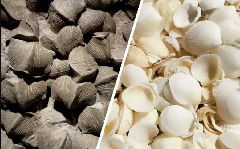
Researchers used Bayesian analysis to study the brachiopods’ decline and bivalves’ rise post-end-Permian extinction, finding bivalves better adapted to changing conditions. Left, Devonian brachiopod fossils from Ohio, USA. Right, recent bivalve shells from Shell Beach, Western Australia. Credit: (Wikimedia Commons; Creative Commons CC0 1.0 Universal Public Domain Dedication) for image on the left. Image on right by Zhong-Qiang Chen.

Researchers used Bayesian analysis to study the brachiopods’ decline and bivalves’ rise post-end-Permian extinction, finding bivalves better adapted to changing conditions. Left, Devonian brachiopod fossils from Ohio, USA. Right, recent bivalve shells from Shell Beach, Western Australia. Credit: (Wikimedia Commons; Creative Commons CC0 1.0 Universal Public Domain Dedication) for image on the left. Image on right by Zhong-Qiang Chen.

One of the biggest crises in Earth’s history, marked by a significant shift in shellfish, saw the widespread replacement of brachiopods, often referred to as ‘lamp shells’, with bivalve species such as oysters and clams. This happened as a result of the devastating end-Permian mass extinction, which effectively reset the evolution of life roughly 250 million years ago.

Research conducted by paleontologists based in Bristol, UK and Wuhan, China has shed new light on this crucial turnover when ocean ecosystems changed from ancient-style to modern-style.

Life on land and in the sea is rich and forms particular ecosystems. In modern oceans, the seabed is dominated by animals such as bivalves, gastropods, corals, crustaceans, and fishes. But these ecosystems all date back to the Triassic when life came back from the brink. During that crisis, only one in twenty species survived, and there has been long debate about how the new ecosystems were constructed and why some groups survived, and others did not.

Brachiopods dominated shelled animals before the extinction, however, bivalves thrived after, better adapting to their new conditions.

“A classic case has been the replacement of brachiopods by bivalves,” explained Zhen Guo at Wuhan and Bristol, who led the project. “Paleontologists used to say that the bivalves were better competitors and so beat the brachiopods somehow during this crisis time. There is no doubt that brachiopods were the major group of shelled animals before the extinction, and bivalves took over after.”

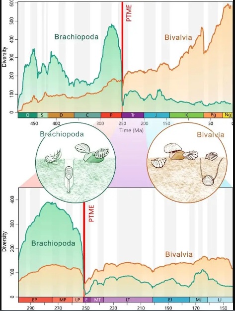
Diversities of brachiopods and bivalves over the past 500 Myr, showing the brachiopod-bivalve switch near the Permian-Triassic boundary. Credit: Zhen Guo et al

Diversities of brachiopods and bivalves over the past 500 Myr, showing the brachiopod-bivalve switch near the Permian-Triassic boundary. Credit: Zhen Guo et al

“We wanted to explore the interactions between brachiopods and bivalves through their long history and especially around the Permian-Triassic handover period,” said Joe Flannery-Sutherland, a collaborator. “So we decided to use a computational method called Bayesian analysis to calculate rates of origination, extinction, and fossil preservation, as well as testing whether the brachiopods and bivalves interacted with each other. For example, did the rise of bivalves cause the decline of brachiopods?”

“We found that in fact, both groups shared similar trends in diversification dynamics right through the crisis time,” said Professor Michael Benton from Bristol’s School of Earth Sciences. “This means that they weren’t really competing or preying on each other, but more probably both responding to similar external drivers such as sea temperature and short-lived crises. But the bivalves eventually prevailed and the brachiopods retreated to deeper waters, where they still occur, but in reduced numbers.”

Professor Zhong-Qiang Chen of Wuhan commented: “It was great to see how modern computational methods can tackle such a long-standing question.

“We always thought that the end-Permian mass extinction marked the end of the brachiopods and that was that. But it seems that both brachiopods and bivalves were hit hard by the crisis, and both recovered in the Triassic, but the bivalves could adapt better to high ocean temperatures. So, this gave them the edge, and after the Jurassic, they just rocketed in numbers, and the brachiopods didn’t do much.”

Zhen Guo said: “I had to check and compile records of over 330000 fossils of brachiopods and bivalves through the study interval, and then run the Bayesian analysis which took weeks and weeks on the Bristol supercomputer. I like the method though because it repeats everything millions of times to take account of all kinds of uncertainties in the data and gives a great deal of rich information about what was going on.”

“The end-Permian mass extinction was the biggest of all time, and it massively reset evolution,’ concluded Professor Benton. “In fact the 50 million years after the crisis, the Triassic, marked a revolution in life on land and in the sea. Understanding just how life could come back from near-annihilation and then set the basis for modern ecosystems is one of the big questions in macroevolution. I’m sure we haven’t said the last word here though!”

Reference: “Bayesian analyses indicate bivalves did not drive the downfall of brachiopods following the Permian-Triassic mass extinction” by Zhen Guo, Joseph T. Flannery-Sutherland, Michael J. Benton, and Zhong-Qiang Chen, 9 September 2023, Nature Communications.
DOI: 10.1038/s41467-023-41358-8

Source: Article By 

@WFS,World Fossil Society, Athira, Riffin T Sajeev,Russel T Sajeev

WFS News: Large fossil spider found in Australia

@WFS,World Fossil Society, Athira, Riffin T Sajeev,Russel T Sajeev

Part (A) and counterpart (B) of Megamonodontium mccluskyi (AM F.145559).

A team of Australian scientists led by Australian Museum (AM) and University of New South Wales (UNSW) paleontologist Dr. Matthew McCurry have formally named and described a fossil spider, Megamonodontium mccluskyi, which is between 11–16 million years old. The findings on this new genus of spider have now been published in the Zoological Journal of the Linnean Society.

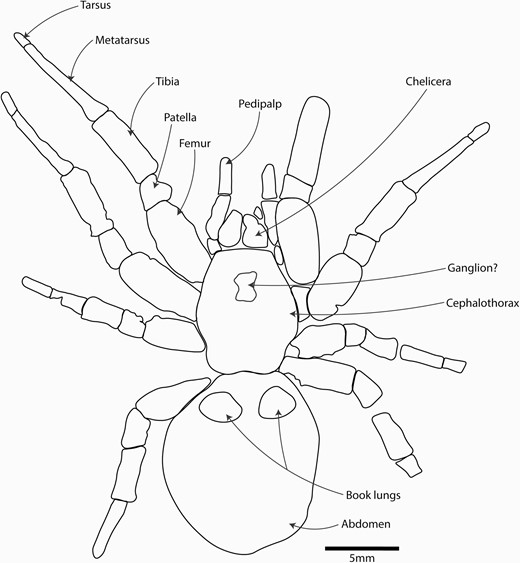
Annotated composite line drawing of AM F.145559, created using the part and counterpart of the fossil.

Annotated composite line drawing of AM F.145559, created using the part and counterpart of the fossil.

Found at McGraths Flat, NSW, a  known for its iron-rich rock called “goethite,” the new genus of spider is the first ever spider  of the Barychelidae family to be found. Similar to the living genus, Monodontium (a brushed trapdoor spider) but five times larger (carapace length, ~10 mm; entire spider, ~50mm from toe to toe), the spider was named after Dr. Simon McClusky who found the specimen. A geospatial scientist based in Canberra, McClusky volunteers his time helping on palaeontological excavations.

Dr. McCurry said that there have been very few fossil spiders found in Australia which makes the discovery very significant.

“Only four spider fossils have ever been found throughout the whole continent, which has made it difficult for scientists to understand their evolutionary history. That is why this discovery is so significant, it reveals new information about the extinction of spiders and fills a gap in our understanding of the past.”

“The closest living relative of this fossil now lives in wet forests in Singapore through to Papua New Guinea. This suggests that the group once occupied similar environments in mainland Australia but have subsequently gone extinct as Australia became more arid.”

Queensland Museum arachnologist, Dr. Robert Raven, who was the supervising author of the study said this was the largest fossil spider to be found in Australia.

“Not only is it the largest fossilized spider to be found in Australia but it is the first fossil of the family Barychelidae that has been found worldwide.”

“There are around 300 species of brush-footed trapdoor spiders alive today, but they don’t seem to become fossils very often. This could be because they spend so much time inside burrows and so aren’t in the right environment to be fossilized.”

University of Canberra Associate Professor, Michael Frese, who used stacking microphotography to scan the fossils said that the fossils from McGraths Flat show an amazing level of detailed preservation.

“Scanning  allowed us to study minute details of the claws and setae on the spider’s pedipalps, legs and the main body. Setae are hair-like structures that can have a range of functions. They can sense chemicals and vibrations, defend the  against attackers and even make sounds.”

The fossil is now housed in the AM’s paleontology collection and is available online for researchers to study.

A separate paper will be published on the same day in the Zoological Journal of the Linnean Society describing a  from McGraths Flat. These are separate publications, but both describe fossils from the same site. Matthew McCurry and Michael Frese are authors on both pieces of work.

More information: Matthew R McCurry et al, A large brush-footed trapdoor spider (Mygalomorphae: Barychelidae) from the Miocene of Australia, Zoological Journal of the Linnean Society (2023). DOI: 10.1093/zoolinnean/zlad100

@WFS,World Fossil Society, Athira, Riffin T Sajeev,Russel T Sajeev

WFS News: The oldest three-dimensionally preserved vertebrate neurocranium.

@WFS,World Fossil Society, Athira, Riffin T Sajeev,Russel T Sajeev

A 455-million-year-old fossil fish provides a new perspective on how vertebrates evolved to protect their brains, a study has found.

In a paper published in Nature today (Wednesday 20th September), researchers from the University of Birmingham, Naturalis Biodiversity Centre in Leiden, Netherlands; and the Natural History Museum have pieced together the skull of Eriptychius americanus.

The research, funded by the Leverhulme Trust, suggests that the ancient jawless fish found in ancient deposits in Colorado, USA has a skull unlike that of any previously seen, and fills a gap currently spanning 100 million years in the evolutionary history of the vertebrate skull.

Using computed tomography, a form of x-ray technique, scientists recreated a detailed 3D representation of the skull of Eriptychius and is the first time that such a comprehensive recreation has been done on the specimen which was collected in the 1940s, originally described in the 1960s and is housed in the Field Museum of Natural History, Chicago.

This ancient fish had separated, independent cartilages encasing the brain, rather than the solid bone or cartilage structure of jawless and jawed fish that followed it.

While later specieshave a fully bound cage of cartilage that holds the brain, these results suggest that the early evolution of structures to separate the brain from other parts of the head may have begun with Eriptychius.

Dr Ivan Sansom, Senior Lecturer in Palaeobiology at the University of Birmingham and senior author of the paper said:

“These are tremendously exciting results that may reveal the early evolutionary history of how primitive vertebrates protected their brains. Eriptychius americanus appears to be the first evidence for a series of cartilages separating the brain from the rest of the head. This study emphasises the importance of museum collections and the application of new techniques in studying them.”

a,b, Photographs of part PF 1795a, which had the split face set in epoxy and was manually prepared (a), and its counterpart PF 1795b, which remains in rock matrix (b). Both are shown in an anatomically ventral view. c,d, Digital model of computed tomographic data of the combined part and counterpart with most of dermal skeleton rendered transparent: anatomical ventral view (corresponding to the visible area of the part in epoxy) (c) and anatomical dorsal view (buried in matrix in the counterpart) (d). Colour scheme for renders: blue-greys, cranial cartilages (matching the detailed scheme in Fig. 2); transparencies, the dermal skeleton; orange, branchial plates; red, orbital plates. Anterior to top in a–d. ant. tess., anterior tesserae; artic. vent. tess., articulated ventral tesserae; branch. plate, branchial plate; cran. cart., cranial cartilages; disp., displaced; frag., fragment; L., left; orb. cart., orbital cartilage; orb. plates, orbital plates; R., right; vasc., vasculature; ?, probable. Scale bar applies to all panels.

a,b, Photographs of part PF 1795a, which had the split face set in epoxy and was manually prepared (a), and its counterpart PF 1795b, which remains in rock matrix (b). Both are shown in an anatomically ventral view. c,d, Digital model of computed tomographic data of the combined part and counterpart with most of dermal skeleton rendered transparent: anatomical ventral view (corresponding to the visible area of the part in epoxy) (c) and anatomical dorsal view (buried in matrix in the counterpart) (d). Colour scheme for renders: blue-greys, cranial cartilages (matching the detailed scheme in Fig. 2); transparencies, the dermal skeleton; orange, branchial plates; red, orbital plates. Anterior to top in a–d. ant. tess., anterior tesserae; artic. vent. tess., articulated ventral tesserae; branch. plate, branchial plate; cran. cart., cranial cartilages; disp., displaced; frag., fragment; L., left; orb. cart., orbital cartilage; orb. plates, orbital plates; R., right; vasc., vasculature; ?, probable. Scale bar applies to all panels.

Dr Richard Dearden, Postdoctoral Research Fellow in Palaeobiology at Naturalis Biodiversity Center and lead author of the paper said:

“On the face of it Eriptychius is not the most beautiful of fossils. However, by using modern imaging techniques we were able to show that it preserves something unique: the oldest three-dimensionally preserved vertebrate head in the fossil record. This fills a major gap in our understanding of the evolution of the skull of all vertebrates, ultimately including humans.”

a–c, Cranial cartilages in estimated life position, with cartilages coloured in pairs in dorsal (a), ventral (b) and anterior (c) view. d,e, Mediolateral cartilages A in dorsal view (d) and median dorsal cartilage in ventral view (e) rendered with a vertical height map texture to emphasize the surface topology. f, Reconstruction of the forebrain relative to the cranial cartilages using a lamprey as a model9,52, shown in dorsal view. g, Cartilages in dorsal view, rendered transparent to show internal vasculature (red). h,i, Cartilages in preserved position in anterior view with dermal skeleton shown (h) and removed (i). Colours in a,b,c,f,h,i as in Fig. 1 with the following additions. Green, dermal skeleton. Red dashed line represents inferred position of mouth in c,h,i. In d and e lighter colours denote areas closer to the camera. Abbreviations as in Fig. 1 with the following additions: antorb. proc, antorbital process; ext. vasc. op., external vascular openings; forebr., forebrain; lat., lateral; medlat. cart, mediolateral cartilage; med. dors. cart, median dorsal cartilage; med., medial; med. vent. cart., median ventral cartilage; med. vent. ridge, median ventral ridge; olf. bulb, olfactory bulb; pin., pineal organ; pin. op., pineal opening; vent., ventral. Scale bar in a is shared by b,c; scale bar in d is shared by e.

a–c, Cranial cartilages in estimated life position, with cartilages coloured in pairs in dorsal (a), ventral (b) and anterior (c) view. d,e, Mediolateral cartilages A in dorsal view (d) and median dorsal cartilage in ventral view (e) rendered with a vertical height map texture to emphasize the surface topology. f, Reconstruction of the forebrain relative to the cranial cartilages using a lamprey as a model9,52, shown in dorsal view. g, Cartilages in dorsal view, rendered transparent to show internal vasculature (red). h,i, Cartilages in preserved position in anterior view with dermal skeleton shown (h) and removed (i). Colours in a,b,c,f,h,i as in Fig. 1 with the following additions. Green, dermal skeleton. Red dashed line represents inferred position of mouth in c,h,i. In d and e lighter colours denote areas closer to the camera. Abbreviations as in Fig. 1 with the following additions: antorb. proc, antorbital process; ext. vasc. op., external vascular openings; forebr., forebrain; lat., lateral; medlat. cart, mediolateral cartilage; med. dors. cart, median dorsal cartilage; med., medial; med. vent. cart., median ventral cartilage; med. vent. ridge, median ventral ridge; olf. bulb, olfactory bulb; pin., pineal organ; pin. op., pineal opening; vent., ventral. Scale bar in a is shared by b,c; scale bar in d is shared by e.

  1. Dearden, R.P., Lanzetti, A., Giles, S. et al. The oldest three-dimensionally preserved vertebrate neurocraniumNature, 2023 DOI: 10.1038/s41586-023-06538-y
University of Birmingham. “Prehistoric fish fills 100 million year gap in evolution of the skull.” ScienceDaily. ScienceDaily, 20 September 2023. <www.sciencedaily.com/releases/2023/09/230920110303.htm>.
@WFS,World Fossil Society, Athira, Riffin T Sajeev,Russel T Sajeev,

WFS News: Rhabdodontidae dinosaurs of Late Cretaceous Europe

@WFS,World Fossil Society, Athira, Riffin T Sajeev,Russel T Sajeev

A new study published in Fossil Record brings together intriguing details about the little-known Rhabdodontidae dinosaurs of Late Cretaceous Europe. These gregarious herbivores, characterized by robust builds and beaks specialized for tough vegetation, inhabited the European archipelago. Despite being widespread and abundant, they vanished in Western Europe due to environmental changes around 69 million years ago, while surviving longer in Eastern Europe. Their fossil record offers valuable insights into their evolution and lifestyle, although its limited nature still challenges comprehensive understanding.

When you think of dinosaurs, you might automatically imagine iconic dinosaurs as Tyrannosaurus and Triceratops. But at the same time when these were stomping on the ancient coastal plains of North America, some of their very distant cousins were reigning over Europe’s lands.

During the Late Cretaceous (between 100 and 66 million years ago), Europe was an extensive archipelago with numerous small and large islands situated in a shallow tropical sea, the so-called Late Cretaceous European Archipelago. The dinosaur groups that lived on these islands were very different from those of other continents, often being much smaller than their mainland relatives. These European dinosaurs include small and medium-sized carnivorous theropods, armoured ankylosaurs, long-necked sauropods, duck-billed hadrosaurs, and rhabdodontids.

Arguably one of the most important of these European dinosaur groups is the family Rhabdodontidae, which groups together the most common medium-sized herbivores of the Late Cretaceous European Archipelago. A joint research team from the Universities of Tübingen (Germany), Budapest (Hungary) and Bucharest (Romania) recently reviewed what we know about these peculiar dinosaurs in a new paper published in the journal Fossil Record.

Generally, rhabdodontid dinosaurs were small to medium-sized animals with an overall body length of approximately 2-6 m. “They were probably habitually bipedal herbivores, characterised by a rather stocky build, with strong hind limbs, short forelimbs, a long tail, and a comparatively large, triangular skull that tapers anteriorly and ends in a narrow snout,” explains Felix Augustin, lead author of the study in Fossil Record.

“They had a relatively robust skull with strong jaws, large teeth and a pointy beak that was covered in keratin, demonstrating that these dinosaurs were well-adapted to eating tough plants.”

In some instances, fossil remains of several individuals of different ages have been found together, indicating that they were gregarious.

Although they died out well before the mass extinction in Western Europe (about 69 million years ago), potentially due to environmental changes that affected the plants they fed on, they survived much longer in Eastern Europe and were among the last non-avian dinosaurs still present before the end of the Cretaceous (66 million years ago).

Interestingly, fossils of rhabdodontids have only been found in Europe and only in rocks ranging in age from 86-66 million years ago, so they were endemic to the Late Cretaceous European Archipelago.

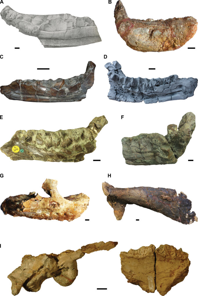
Type specimens of the nine rhabdodontid species described so far. A. The original drawing of the lectotype of Rhabdodon priscus, MPLM 30, a partial left dentary. The specimen has since deteriorated (Pincemaille-Quillevere 2002). Modified after Matheron (1869). B. Holotype of Rhabdodon septimanicus, MDE D-30, an incomplete right dentary. Photo kindly provided by Eric Buffetaut. C. Lectotype of Mochlodon suessi, PIUW 2349/2, a right dentary. D. Holotype of Mochlodon vorosi, MTM V 2010.105.1, a left dentary. E. Holotype of Zalmoxes robustus, NHMUK R.3392, a right dentary. Photo kindly provided by János Magyar. F. Holotype right dentary of Zalmoxes shqiperorum, NHMUK R.4900. Note that the holotype of Z. shqiperorum also comprises several postcranial elements that presumably belong to the same individual as the dentary. Photo kindly provided by János Magyar. G. Holotype of Matheronodon provincialis, MMS/VBN-02-102, a right maxilla. Modified after Godefroit et al. (2017). H. Holotype of Pareisactus evrostos, MCD 5371, a left scapula. Modified after Párraga and Prieto-Márquez (2019). I. Holotype of Transylvanosaurus platycephalus, LPB (FGGUB) R.2070, a partial skull comprising the articulated basicranium and both frontals. Scale bars: 1 cm.

Type specimens of the nine rhabdodontid species described so far. A. The original drawing of the lectotype of Rhabdodon priscus, MPLM 30, a partial left dentary. The specimen has since deteriorated (Pincemaille-Quillevere 2002). Modified after Matheron (1869). B. Holotype of Rhabdodon septimanicus, MDE D-30, an incomplete right dentary. Photo kindly provided by Eric Buffetaut. C. Lectotype of Mochlodon suessi, PIUW 2349/2, a right dentary. D. Holotype of Mochlodon vorosi, MTM V 2010.105.1, a left dentary. E. Holotype of Zalmoxes robustus, NHMUK R.3392, a right dentary. Photo kindly provided by János Magyar. F. Holotype right dentary of Zalmoxes shqiperorum, NHMUK R.4900. Note that the holotype of Z. shqiperorum also comprises several postcranial elements that presumably belong to the same individual as the dentary. Photo kindly provided by János Magyar. G. Holotype of Matheronodon provincialis, MMS/VBN-02-102, a right maxilla. Modified after Godefroit et al. (2017). H. Holotype of Pareisactus evrostos, MCD 5371, a left scapula. Modified after Párraga and Prieto-Márquez (2019). I. Holotype of Transylvanosaurus platycephalus, LPB (FGGUB) R.2070, a partial skull comprising the articulated basicranium and both frontals. Scale bars: 1 cm.

The group currently comprises nine different species from five European countries (France, Spain, Austria, Hungary, and Romania).

“The first rhabdodontid species was scientifically named more than 150 years ago and the last one as recently as November 2022, so, although the group looks back to a long research history, we still have much to learn about it,” says Felix Augustin.

“Generally, our portraying of the world of dinosaurs is heavily biased towards the well-known North-American and Asian dinosaur faunas,” he adds.

Dinosaur fossils from the Late Cretaceous are much rarer in Europe than in North America or Asia, and thus far no complete skeleton of a rhabdodontid has been described. Even though they were so abundant and common in the Upper Cretaceous of Europe, several key aspects about them remain poorly known, including their detailed body proportions, their posture and locomotion, as well as their feeding behaviour.

“In the past decades, a wealth of new, and often well-preserved, rhabdodontid fossils has been discovered throughout Europe, the majority of which still remains to be studied,” says Felix Augustin. “A joint research project is currently underway to study the available fossil material in order to gain new insights into the evolution and lifestyle of these fascinating yet still poorly known dinosaurs.”

  1. Felix J. Augustin, Attila Ősi, Zoltán Csiki-Sava. The Rhabdodontidae (Dinosauria, Ornithischia), an enigmatic dinosaur group endemic to the Late Cretaceous European ArchipelagoFossil Record, 2023; 26 (2): 171 DOI: 10.3897/fr.26.108967
Pensoft Publishers. “Europe’s very own dinosaurs — the enigmatic Late Cretaceous rhabdodontids.” ScienceDaily. ScienceDaily, 1 September 2023. <www.sciencedaily.com/releases/2023/08/230830131713.htm>.
@WFS,World Fossil Society, Athira, Riffin T Sajeev,Russel T Sajeev

WFS News: Sulfur minerals that make fossils are especially well-suited to radiography

@WFS,World Fossil Society, Athira, Riffin T Sajeev,Russel T Sajeev

New research reveals that sulfur minerals that make fossils in the Norwegian archipelago are especially well-suited to radiography.

An overlook of the Muen plateau as seen from the Muen mountain on Edgeøya, Svalbard. Marine reptiles are spread out across the plateau. Author VSE in red jacket for scale. Credit: Sofie Bernhardsen, CC-BY 4.0

An overlook of the Muen plateau as seen from the Muen mountain on Edgeøya, Svalbard. Marine reptiles are spread out across the plateau. Author VSE in red jacket for scale. Credit: Sofie Bernhardsen, CC-BY 4.0

X-ray analysis has led to the categorization of a previously-unidentified marine reptile fossil discovered in Edgeøya, Svalbard. The research was recently published in the journal PLOS ONE.  The study, conducted by Victoria S. Engelschiøn of the University of Oslo and her team, suggests that this method could reveal fresh insights about ancient life in the future.

The effectiveness of X-ray techniques in investigating well-conserved fossil remains is often dependent on the condition of preservation, which can vary greatly across different sites. Through this study, Engelschiøn and her team showed that fossils from the Middle Triassic Botneheia Formation in Svalbard, Norway, are particularly suitable for radiographic imaging.

The focus of this study is a fossil marine reptile whose remains are compressed and encased in shale. It lived around 240 million years ago, when Svalbard was covered by an ocean. After it died, it sank to the seafloor and was buried in the mud, then became extremely flattened over time. Originally excavated in 2008, the identity of this fossil has since been debated. X-ray imaging of the specimen revealed new details, including features of the skull and teeth that allowed researchers to conclude that this reptile most likely belongs to the ichthyosaur species Phalarodon atavus.

Photograph of specimen PMO 219.250. Credit: Engelschiøn et al. PLOS ONE, CC-BY 4.0

Photograph of specimen PMO 219.250. Credit: Engelschiøn et al. PLOS ONE, CC-BY 4.0

The authors also examined the mineralogy of fossils from this formation, identifying multiple forms of sulfate minerals, notably including sulfate baryte, which gives the fossils very high X-ray contrast, allowing for the high quality of radiographic imaging. The formation of these minerals is little understood but could be linked to conditions created by ancient volcanic activity. Thus, this study not only demonstrates the utility of X-ray techniques for studying these fossils, but also identifies conditions that can form fossils well-suited for these techniques, in Svalbard and potentially elsewhere.

The authors add: “The rocks from Svalbard are full of flattened marine reptiles. Our discovery of the exceptional X-Ray contrast means that we can learn much more about these ancient predators than we previously thought.”

Reference: “Exceptional X-Ray contrast: Radiography imaging of a Middle Triassic mixosaurid from Svalbard” by Victoria S. Engelschiøn, Aubrey J. Roberts, Ruben With and Øyvind Hammer, 31 May 2023, PLOS ONE.
DOI: 10.1371/journal.pone.0285939

@WFS,World Fossil Society, Athira, Riffin T Sajeev,Russel T Sajeev