Images of the three-horned dinosaur Triceratops battling with conspecifics or the predatorTyrannosaurus have become ingrained in both the scientific and the popular mind. Lesions (wounded or diseased areas) on the horns, frill, and face of Triceratops specimens have been cited as evidence in support of the defensive and offensive nature of the animal’s cranial ornamentation . An alternative interpretation posits that these structures functioned in visual display rather than combat. To date, discussions of osteopathology in Triceratops have been anecdotal, focusing on generating speculative scenarios to explain instances of hypothesized injury . Without a rigorous statistical analysis, however, it is impossible to relate injury patterns to specific behaviors.
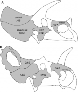
We surveyed cranial specimens from adult individuals of the ceratopsid dinosaurs Triceratops andCentrosaurus for bony lesions . The two animals differ greatly in cranial ornamentation; Triceratops has two large supraorbital horncores and a smaller nasal horncore, whereas Centrosaurus has a large nasal horncore and a pair of small supraorbital horncores (Figure 1).

Schematics of the skulls of (A) Triceratops and (B) Centrosaurus, showing incidence rates of lesions (periosteal reactive bone and fracture calluses) on each cranial element (number of abnormal elements / total number of elements). Not to scale.
In modern horned animals, the morphology and location of the horns is closely associated with combat styles . By analogy, it is then expected that if Centrosaurus and Triceratops engaged in horned combat with conspecifics, the two genera would have had very different forms of combat. Thus, relative rates of lesion occurrence should differ between comparable cranial elements in both genera. If cranial ornamentations were used exclusively for visual display and/or species recognition, and not for physical contact, the two taxa are predicted to have similar rates of incidence for cranial lesions in all comparable cranial elements.
Results
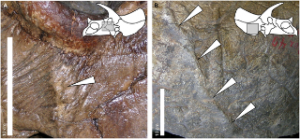
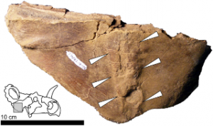
Cranial abnormalities observed in both taxa included periosteal reactive bone, healed and healing fractures, and resorptive bone lesions of unknown etiology (Figures 2,3). Only the first two categories, considered most likely due to trauma , were included in further statistical analysis .
Periosteal reactive bone reflects superficial trauma; the reaction is caused by separation of the periosteum from underlying layers and subsequent inflammatory response and healing of the bone. Evidence of this injury presents as an elevated, remodeled ridge on the external surface of the bone, which may cut across the normal pattern of neurovascular impressions on the surface of the skull (Figure 2). Periosteal reactive bone was the most common of the observed pathologies (22 out of 26 observed lesions considered here).
Calluses associated with healed or healing fractures constitute the second variety of observed lesions (Figure 3; 4 out of 26 observed lesions). Such features result from the several steps of bone growth intended to reunify mechanically or pathologically separated pieces of bone. The process progresses from a primary callus with disorganized bone to a secondary callus of secondary bone . Because primary bone can be preserved, calluses can be discovered in different stages of healing. The character and appearance of calluses is difficult to predict as the proliferation of bone at the site can vary from minimal to extremely exuberant depending on the individual and the severity of the fracture. In the fossil record, the fractures and calluses are often associated with an overall displacement of the bone that extends for a considerable area. Typically, fractures present as a full-thickness feature in the bone with observed disturbances of the bone fabric on both the medial and lateral aspects of the bone. This contrasts with instances of periosteal reactive bone, which affect only one side of the element.
Statistical Analysis
A G-test of independence was used for all comparisons . Triceratops and Centrosaurus did not differ significantly in the rates of lesion occurrence within the nasal, jugal, or parietal bones of the skull (P>0.20 in all cases; present full data). In contrast, Triceratops had significantly higher prevalence of lesions on the squamosal bone of the frill than did Centrosaurus (P = 0.002; see Figure 1 and table S1 for full data, and Figure 3 for the sole pathological squamosal fromCentrosaurus).
Discussions
We reject the possibility that a generalized pathogenic factor (such as a habitat-specific fungal infection) caused the differing prevalence of lesions between Triceratops and Centrosaurus, because all cranial elements should then show similar rates of incidence. We also rule out predatory attacks as the primary cause of the lesions, because similar large predators (tyrannosaurid theropods) were present in the habitats for both genera, and we would thus expect similar patterns of osteological abnormalities in both. Alternatively, it might be claimed that Triceratops had more frequent occurrence of lesions on the squamosal because this element forms a greater proportion of the frill’s exposed area, and was thus more likely to be injured, than in Centrosaurus (e.g., Figure 1). We tested this hypothesis by comparing the prevalence of lesions in the entire frill of both taxa and still found a significant difference between the two (3 pathological and 84 non-pathological specimens forCentrosaurus; 10 pathological and 59 non-pathological specimens for Triceratops; P = 0.012; for full explanation). Instead, the evidence appears to be most consistent with the majority of cranial abnormalities in Triceratops being generated by the horns of conspecifics. The observed instances of periosteal reactive bone and healing fractures are consistent with such non-random trauma, and the elevated rates of abnormal bone morphology within the frill bones are consistent with predictions from modeling of horn-to-horn combat This suggests that the cranial ornamentation of ceratopsids, particularly Triceratops, was not only for visual display but that the horns also had a real role in physical combat.
It is important to note that we do not claim to infer a precise cause for individual pathologies on certain specimens (that a slip of a horn during a specific bout caused the injury to the jugal in YPM 1822, for example; Figure 2A). Certainly, at least some of the pathologies noted here may not be due to combat. We only claim that the overall pattern in all of the specimens is consistent with intraspecific combat in Triceratops.
Non-ceratopsid neoceratopsians (e.g., Protoceratops), the evolutionary predecessors of ceratopsids, possessed a thin, enlarged frill but lacked elongated brow or nasal horns. Thus, the primitive function of the frill (in addition to a role in jaw muscle attachment) was probably that of display rather than cervical protection . The later evolution of brow horns would have increased the importance of a protective function for the frill, assuming that the horns were used in combat. The relatively thickened, solid frill of Triceratops may have been an exaptation for cervical protection, in addition to a role in display. This suggests interesting possibilities for the factors that drove the evolution of cranial morphology in ceratopsids. Display probably was an important function for the horns and frills in all ceratopsids, but not the only one. Horned combat, and the consequences of injury from this combat, may have been another important selective factor. Recent discoveries strongly suggest thatCentrosaurus evolved from an ancestor with a Triceratops-like horn configuration . One evolutionary interpretation worthy of further consideration is that some ceratopsids (such asCentrosaurus) lost their long brow horns or changed combat styles as a way to reduce cranial injury. This interpretation also suggests that the frill may not have had a protective function withinCentrosaurus (as evidenced by the reduced occurrence of lesions on the squamosal, relative toTriceratops), but instead functioned for species recognition and/or other forms of visual display.Centrosaurus and some other ceratopsids may have focused blows on an opponent’s torso rather than the skull; this is suggested by the occurrence of fractured ribs in Centrosaurus, Pachyrhinosaurus, and Chasmosaurus . Statistical analysis and comparison with rates of rib fracture in Triceratops, as well as rates of cranial bony anomalies in additional taxa, may be informative in further evaluating this hypothesis. Clearly, horned dinosaurs used their cranial ornamentations for a variety of functions.
Andrew A. Farke1*, Ewan D. S. Wolff2, Darren H. Tanke3
1 Raymond M. Alf Museum of Paleontology, Claremont, California, United States of America, 2 Department of Pathobiological Sciences, School of Veterinary Medicine, University of Wisconsin-Madison, Madison, Wisconsin, United States of America, 3 Royal Tyrrell Museum of Palaeontology, Drumheller, Alberta, Canada


 September 23rd, 2012
  Riffin

 Posted in
  Tags:
Tags: 