@ WFS,World Fossil Society,Riffin T Sajeev,Russel T Sajeev
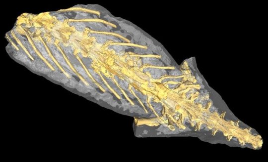
Synchrotron tomography permitted access to the inside of the “mummy”. The skeleton and several organs are perfectly preserved.Credit: Jérémy Tissier; CC BY
A new study on an exceptionally preserved salamander from the Eocene of France reveals that its soft organs are conserved under its skin and bones. Organs preserved in three dimensions include the lung, nerves, gut, and within it, the last meal of the animal, according to a study published in the peer-reviewed journal PeerJ by a team of palaeontologists from France and Switzerland.
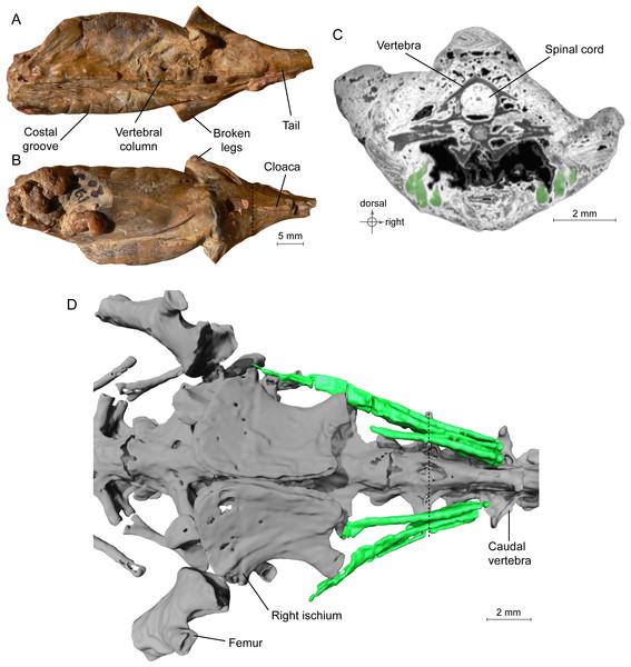
Specimen MNHN.F.QU17755, holotype of Phosphotriton sigei.
(A and B) Fossil in dorsal and ventral views. Some characteristics of urodeles, such as costal grooves or scaleless skin, are observable on the external aspect of the specimen. The cloaca and vertebral column are visible. The dotted line represents the position of the tomogram illustrated in Fig. 1C. (C) Tomogram of the tail part of the animal showing the muscles, in green, ventral and lateral to the vertebrae, and the spinal cord preserved inside the neural canal of a vertebra. Bony material is characterized by a dark grey shade, because of its light density, compared to the mineral matrix (grey or white) and void (black). Soft-tissues are also mostly darker than the mineral matrix, but are mainly recognizable by their structure and shape, on tomograms or in 3D. (D) 3D reconstruction of undetermined tail muscles, in green, which could attach to the ischium or femur. Dotted line represents the position of the tomogram illustrated in Fig. 1C.
Accessing the complete anatomy of an extinct animal, i.e. both its external and internal aspects, has often been the dream of palaeontologists. Indeed, in 99% of cases, fossils are only represented by hard parts: bones, shells, etc. Fossils preserving soft tissues exist, but they are extremely rare. However, their significance for science is enormous. What did the animal look like? What did they eat? How did they live? Most of these questions can be answered by exceptionally preserved fossils.
The newly studied fossil externally looks like a present-day salamander, but it is made of stone. This fossil “mummy” is the only known specimen of Phosphotriton sigei, a 40-35 million years old salamander and belongs to the same family as the famous living fire salamander (Salamandra salamandra).
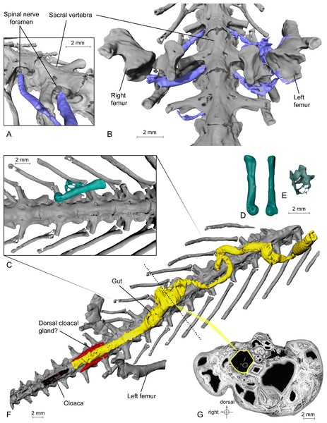
Exceptional preservation of nerves, digestive tract and stomachal content.
(A and B) 3D reconstructions of the pelvic section of MNHN.F.QU17755, in laterodorsal (A) and ventral (B) views. The lumbosacral plexus (in blue) is partly preserved. Nerves exit the last trunk, the sacral and the first caudosacral vertebrae through the spinal nerve foramina. (C) Preserved bones of an anuran frog (ranoid?), in green, inside the digestive tract (not shown, to better reveal its content; see Fig. 3F) of MNHN.F.QU17755. (D) Anuran humerus found inside digestive tract of MNHN.F.QU17755, in lateral and ventral views. (E) Anuran vertebrae found inside digestive tract of MNHN.F.QU17755. The centrum is very thin; the holes may represent segmentation artifacts. (F) 3D reconstruction of MNHN.F.QU17755 in ventral view, showing the nearly complete digestive tract. The caudal end is very close to the cloaca, and is bordered near the pelvic girdle by presumed dorsal cloacal glands (see Fig. 4A). The dotted line represents the position of the virtual section illustrated in Fig. 3G. (G) Virtual section of the trunk, showing the digestive tract (in yellow) and its content (frog bones).
It is unfortunately incomplete: only the trunk, hip and part of hind legs and tail are preserved. Until very recently, the only thing palaeontologists could tell about this specimen was visible anatomical details, such as the cloaca, the orifice used for reproduction and by digestive and urinary canals. Indeed, though it was discovered in the 1870s, it was never studied in detail.
Thanks to recent synchrotron technology, its skeleton1 and various organs2 could be studied. The specimen was scanned at the ID19 beamline of the European Synchrotron Radiation Facility (ESRF) in Grenoble (France). This modern technology gave access to an incredible level of details that could never have been achieved before without slicing the specimen into a series of thin sections.
The quality of preservation is such that looking at the tomograms (equivalent of radiograms) feels like going through an animal in the flesh. At least six kinds of organs are preserved in almost perfect condition, in addition to the skin and skeleton: muscles, lung, spinal cord, digestive tract, nerves, and glands.
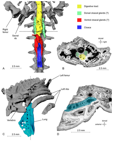
Exceptional preservation of cloacal glands (?) and lung.
(A) 3D reconstruction of supposed dorsal and ventral cloacal glands, in ventral view, under the two ischia (not shown). The dorsal cloacal glands are located between the first and second caudosacral vertebrae and the digestive tract (see Fig. 4B). The ventral cloacal glands are located under the digestive tract and anterodorsal to the cloaca. The dotted line represents the position of the virtual section illustrated in Fig. 4B. (B) Virtual section of the pelvic girdle, illustrating the digestive tract and the dorsal cloacal glands, between a caudal vertebra and the two ischia. (C) 3D reconstruction of the incomplete lung (in blue), inside the specimen MNHN.F.QU17755, in oblique anterior view. It is located lateroventrally to the trunk vertebrae, in the anteriormost preserved part of the fossil. The dotted line represents the position of the tomogram illustrated in Fig. 4D. (D) Virtual section of the anteriormost preserved part of MNHN.F.QU17755, showing the inside of the lung in lateral view.
But the most incredible is the preservation of frog bones within the stomach of the salamander. Salamanders almost never eat frogs or other salamanders, though they are known to be quite opportunistic. Was it a last resort meal or a customary choice for this species? This, unfortunately, will probably never be known.
These new results are described by Jérémy Tissier from the Jurassica Museum and the University of Fribourg in Switzerland, and Jean-Claud Rage and Michel Laurin, both from the CNRS/Museum national d’histoire naturelle/UPMC in Paris.
Author Michel Laurin notes, “This fossil, along with a few others from the same lost site, is the most incredibly well-preserved that I have seen in my entire career. And now, 140 years after its discovery, and 35 million years after the animal died, we can finally study it, thanks to modern technology. The mummy returns!”
@ WFS,World Fossil Society,Riffin T Sajeev,Russel T Sajeev
- Jérémy Tissier, Jean-Claude Rage, Michel Laurin. Exceptional soft tissues preservation in a mummified frog-eating Eocene salamander. PeerJ, 2017; 5: e3861 DOI: 10.7717/peerj.3861
- Source: ScienceDaily, 3 October 2017. <www.sciencedaily.com/releases/2017/10/171003093929.htm


 October 4th, 2017
  Riffin
  Posted in
  Tags:
Tags: 