@WFS,World Fossil Society,Riffin T Sajeev,Russel T Sajeev
“The respiratory organs of vertebrates exhibit a tremendous degree of diversity, but the lung-air sac system of birds is truly unique among extant species,” says Dr. Markus Lambertz from the Institute for Zoology at the University of Bonn in Germany. Air sacs are bellows-like protrusions of the lung, and their volume changes cause the air flow in the separate gas exchanger. This functional separation is crucial for the exceptional efficiency of this respiratory system, but air sacs can do more: they can invade bones, a process called “pneumatization.”
Pneumatized bones are very light, because they are filled with air instead of the more heavy marrow, which was not only important for active flight, but also for the evolution of gigantism in sauropod dinosaurs. Through the presence of the resulting pneumatic cavities, it has long been known that air sac-like structures predate the origin of birds, since they were found both in the gigantic sauropods as well as in carnivorous dinosaurs. However, when and potentially how many times air sacs did evolve was inaccessible until now.
Pneumosteum: a hitherto unknown type of bony tissue as a diagnostic tool
Filippo Bertozzo was pretty surprised when he analyzed the bone structure in the course of his master’s thesis at the Steinmann-Institute for Geology, Mineralogy and Paleontology of the University of Bonn: “Bones that are in contact with air sacs exhibit a unique structure composed of very fine and densely packed fibers. After it turned out that this was true both in modern birds and extinct dinosaurs, we proposed to name this special kind of bony tissue “pneumosteum.” ”
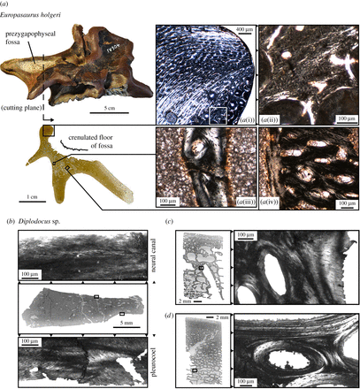
Especially astonishing was the fact that pneumosteum was not only restricted to pneumatized bones, but was also found on the surface of conspicuous cavities present in cervical vertebrae of sauropod dinosaurs. Dr. Lambertz adds: “Such cavities had already previously been hypothesized as potential locations of air sacs, but only our microscopic analysis now provides convincing arguments for this.”

Pneumosteum in sauropods. (a) Cross section of left prezygapophysis of Europasaurus holgeri cervical vertebra. The lamina connecting neural spine and prezygapophysis consists of primary fibrolamellar bone dorsally (a(i)) and metaplastic bone ventrally (a(ii)). The crenulated surface of the lateral fossa is composed of secondary trabecular bone showing densely packed fine fibres: pneumosteal bone (a(iii–iv)). The appearance of pneumosteal fibres is markedly different from the coarse Sharpey’s fibres associated with tendinous insertions a(ii). (b) Pneumatized vertebrae of Diplodocus sp. present pneumosteal bone facing the pleurocoel, but not the floor of the neural canal, which is lined by parallel-fibred bone. (c) Pneumatized anterior caudal vertebrae exhibit pneumosteum in their loose trabeculae. (d) Apneumatic middle caudal vertebrae do not exhibit pneumosteum in their denser trabeculae consisting of regular lamellar bone. (Online version in colour.)
Other soft tissues, such as muscles, can leave traces in bone as well. “There are several types of fibers within bone tissue, but the pneumosteum is markedly different from them,” explains Prof. Dr. Martin Sander from the Steinmann-Institute in Bonn. This characteristic individuality of the pneumosteum thus makes it an excellent diagnostic tool for recognizing bones that were in contact with air sacs.

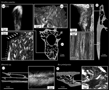
Pneumosteum in birds and its absence in mammals. (a) Ostrich cervical vertebra cut transversely revealing Sharpey’s fibres at tendinous insertions a(i) and pneumosteum in air-sac-associated secondary endosteal trabeculae (a(ii–iii)). The right part of the centrum cortex consists of primary fibrolamellar bone whereas the left lower part of pneumosteum (a(iii), asterisks), truncated by an endosteal resorption front (arrows). (b) Longitudinal section of cervical rib with pneumosteal trabeculae proximally (b(i)) and ossified tendons distally (b(ii)). (c) Subfossil moa sternum exhibiting pneumosteum in the secondary trabeculae. (d) The pneumatized skull of the aurochs lacks pneumosteum. (Online version in colour.)
Access to the past and potential for future research
Given that pneumosteum was only discovered in the dinosaurian lineage now provides the opportunity to trace the evolutionary origin of air sacs. Especially the fact that pneumosteum is not restricted to pneumatized bones but was also found on bone surfaces opens up access to studying species that might have exhibited air sacs as part of their respiratory system, but lack obviously pneumatized bones.
Fossilization of air sacs is nearly impossible because their delicate structure is composed of only a few layers of cells. Professor Sander thus is convinced that the discovery of pneumosteum will lead to a greatly improved understanding of the evolution of the dinosaurian respiratory system. Dr. Lambertz concludes with: “This project once again highlights the importance of the interdisciplinary collaboration between zoologists and paleontologists for elucidating evolutionary history.”
- Markus Lambertz, Filippo Bertozzo, P. Martin Sander. Bone histological correlates for air sacs and their implications for understanding the origin of the dinosaurian respiratory system. Biology Letters, 2018; 14 (1): 20170514 DOI: 10.1098/rsbl.2017.0514


 January 4th, 2018
  Riffin
  Posted in
  Tags:
Tags: 