@ WFS,World Fossil Society,Riffin T Sajeev,Russel T Sajeev
Citation: Das AJ, Murmann DC, Cohrn K, Raskar R (2017) A method for rapid 3D scanning and replication of large paleontological specimens. PLoS ONE 12(7): e0179264. https://doi.org/10.1371/journal.pone.0179264
Editor: Pasquale Raia, Seconda Universita degli Studi di Napoli, ITALY

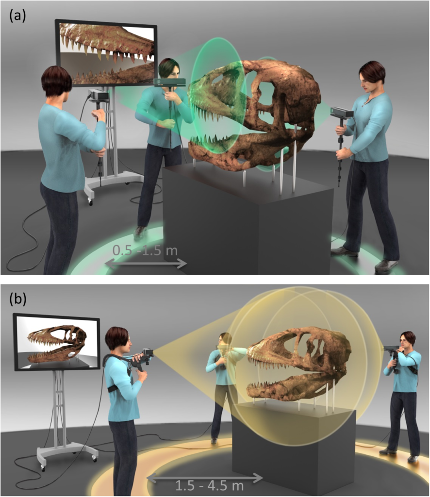
Scanning technique.(a) Small section, high resolution scanning. The user holds a monopod mounted Kinect at close range (0.5–1.5 m) from the target. (b) Large section or complete 360° scanning. The user mounts Kinect on a body supported rig and walks around the artifact (1.5–4.5 m from target) to complete the scan. Sketch by Francis Goeltner.
The field of paleontology has transformed in the last few years as a result of the developments in 3D scanning technology and rendering software that have enhanced the quality of virtual models [1–4]. Conventionally, a photograph is utilized for research purposes which has its benefits but also has limited application. A two dimensional (2D) image is easy to capture, interpret and is still a useful method of analysis in paleontology research [5–7]. However, a 2D image cannot capture the details regarding depth of the scene. Recent studies have shown that 3D scanning and analysis of specimens can provide rich information which can be beneficial in a range of studies [8]. These techniques are increasingly seen in museums and research labs due to the compact nature of some of the imaging devices [3, 4]. 3D scanning can provide depth maps in a non-invasive, non-contact manner which is attractive for studying paleontological specimens due to their delicate physical properties. For instance, it has been used to estimate the mass of dinosaurs by combining it with computer modeling [9]. It has also been used to create virtual skeletons for different fauna for comparative purposes [10]. Other examples of 3D scanning in related fields include typology [11], pottery studies [12] and footprint analysis in archaeology [13].
At the heart of 3D imaging technology is the 3D scanner itself. There are several approaches to perform 3D scanning from structured light scanners to computed tomography (CT). However, most of these scanners are industrial or clinical grade instruments and are generally very expensive and bulky. Structured light scanners need calibration and are inherently expensive due to the requirement of a laser projector and a high end camera to capture the images. There are reports of using structured light based 3D scanning for fossils of the size of several tens of centimeters [14] but not large specimens like T.rex skulls [10]. Several other reports have demonstrated the use of CT imaging due to its ability to study internal details of specimens. However, CT scanners are expensive and the imaging is done at a clinical facility [15, 16]. Additionally, most studies have used these techniques on small specimens due to the complexity of the scanner and also restriction of the data size that can be handled by the software for large specimens. For instance, a high resolution dental scanner would not be able to handle the large data size when scanning the jaw of a T.rex. Hence, there are limitations in the volume of the object that can be scanned with these methods, the ease of setup and processing the data. Furthermore, the software for these industrial scanners is proprietary making it inaccessible to researchers and museums. Although there have been some reports on the use of free open source photogrammetric software for 3D imaging, the process is cumbersome requiring a large amount of data to reconstruct the models [17]. Hence there is a need for a technique that is accurate, low-cost, easy to implement, has open source software capability and can be adapted for large scale paleontological scanning.
We propose a new technique that provides high quality 3D reconstructions of large specimens with relative ease. We used the Kinect v2 TOF sensor to perform 3D scanning of large paleontological specimens for the first time. Kinect has traditionally been used in gesture recognition [18–20] in gaming, computer graphics [21] and more recently in 3D scanning [22–24]. There has been one earlier report that used Kinect v1 for paleontological specimens but the reconstructions were noisy and smoothing the data resulted in loss of features [14]. Kinect v1 uses structured light imaging in contrast to Kinect v2 that is based on TOF imaging which has significantly improved since the report by Falkingham [14]. The sensor technology in Kinect v2 is not only superior to Kinect v1 but also the computation aspect has improved providing real-time high quality reconstructions. The Kinect has shown to be a promising tool for full body scanning with improvements in registration and alignment techniques [25]. Most of the earlier demonstrations have been performed on rotating objects where the Kinect is stationary. However, this may not be possible for large paleontological specimens that are housed in enclosures that cannot be modified.
In this report, we present a method for 3D scanning that is well suited for paleontology and has the following advantages; a) It has an short acquisition time of 60-120s even for large specimens, b) Since the scanner is compact so it can be moved around the specimen on a tripod or adapted to a body-mounted wearable geometry; c) The entire set-up being low-cost and the availability of free scanning and post-processing software.
@ WFS,World Fossil Society,Riffin T Sajeev,Russel T Sajeev


 April 1st, 2018
  Riffin
  Posted in
  Tags:
Tags: 