 November 12th, 2013
  riffin
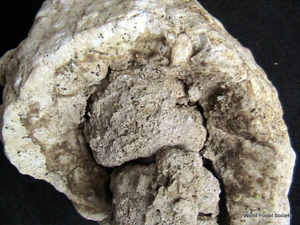
 By integrating high-resolution X-ray imaging (termed microCT), 3D image segmentation, and computer animation, a new study conducted by Carole Gee at the University of Bonn, Germany, demonstrates the visualization of fossils without destroying the material. Traditional techniques, such as thin-sectioning, require investigators to physically cut up the fossil in order to observe internal structures. Dr. Gee, however, has now successfully applied microCT to visualize silicified conifer seed cones as old as 150 million years without cutting, sawing, or damaging the specimens in any way.
Well-preserved, informative plant fossils are few and far between. Specimens with reproductive organs are especially scarce but are invaluable to understanding plant evolution and ancient diversity. When such fossils are unearthed, they are lucky finds and often only single specimens are present.
“Because each specimen is precious, the main goal of this research was to study the internal structure of fossil conifer seed cones without destroying or damaging them,” explains Gee.
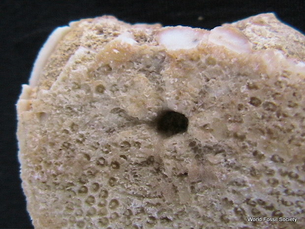
Using this technique, X-ray images, similar to those used in the medical field, are captured, providing virtual cross-sections of the specimen, without ever cutting into the sample. These images are then combined, producing a 3D reconstruction. This study, along with computer animations and detailed figures presenting microCT imaging, is freely available for viewing in the November issue of Applications in Plant Sciences.
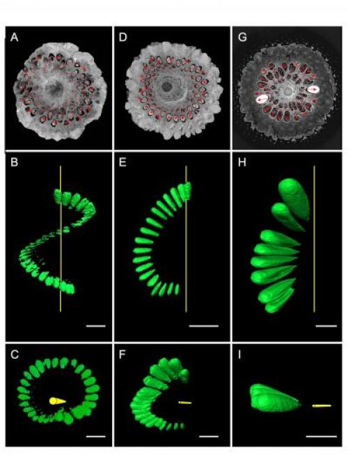
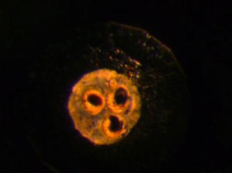
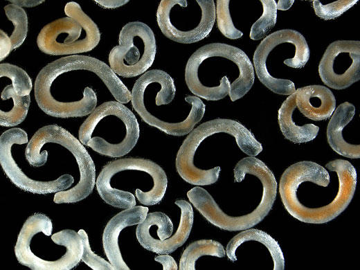
This shows fossil and recent araucarian cones sectioned in 2D by microCT (A, D, G), and showing one segmented spiral or row of seeds or seed locules produced by 3D imaging (B, C, E, F, H, I). The seed spirals or rows in A, D, and G are delineated by red arrows. Yellow lines in B, C, E, F, H, and I represent the polar axis through the cones. Scale bars = 1 cm. (A–C) Fossil cone of Araucaria sp. from Wyoming (specimen no. CG066, Flynn Collection). (A) Transverse section 294/1012; diameter = ca. 6 cm. (B) Lateral view showing the 360° revolution of a single seed spiral. (C) Oblique distal view. (D–F) Fossil cone of Araucaria mirabilis from the Middle Jurassic of Argentina (specimen no. K5640, Museum für Naturkunde Chemnitz collection). (D) Transverse section 280/933; diameter = ca. 7.5 cm. (E) Lateral view showing the 180° revolution of a single seed spiral. (F) Oblique distal view. (G–I) Recent cone of Araucaria araucana from the Economic Botany Garden, University of Bonn, Germany. (G) Transverse section 469/876; diameter = ca. 17 cm. (H) Lateral view showing the vertical (nonspiral) arrangement of a row of seeds. (I) Oblique distal view. (Credit: Image credit Carole T. Gee. From Gee, C. T. 2013. Applying microCT and 3D visualization to Jurassic silicified conifer seed cones: A virtual advantage over thin-sectioning. Applications in Plant Sciences 1(11): 1300039. doi:10.3732/apps.1300039.)
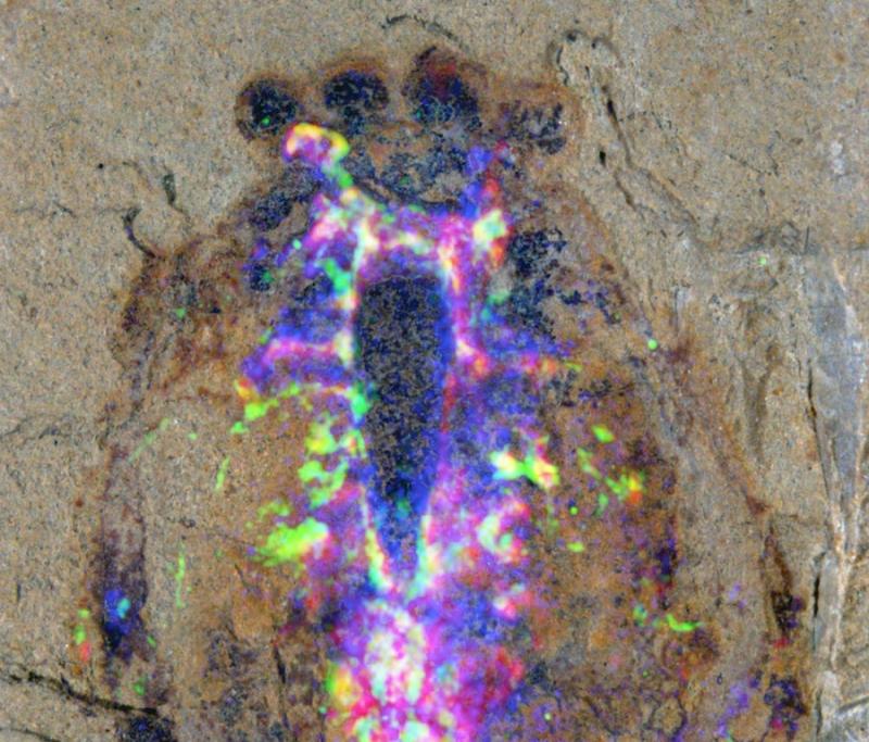
In the study, Gee demonstrates how this technique allows the observation of internal features such as seeds, vascular tissue, and cone scales. Furthermore, by adding artificial color to highlight certain structures or tissues, such as a row of seeds within a cone, the natural pattern of growth was evident. As Gee observes, “It’s amazing to visualize internal structures of dinosaur-aged fossils in such great detail without cutting up the fossil or damaging them at all.”
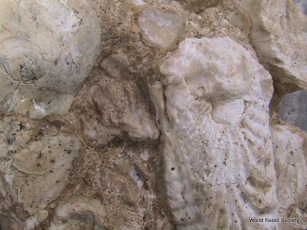
It was then possible to identify the fossils as belonging to three distinct families: Pinaceae — the pine family, Araucariaceae — a family of coniferous trees currently found only in the Southern Hemisphere, and Cheirolepidiaceae — a now-extinct family of conifers known only from the Mesozoic.
“This tells us that 150 million years ago, the ancient forests of western North America consisted of members of these three families. The fossil cones of the Araucariaceae from Utah confirm that this family, which now grows naturally in Australasia and South America, once had a worldwide distribution,” notes Gee.
Dr. Gee hopes this study will provide researchers with an alternative to traditional techniques such as thin-sectioning, which often leave the fossil completely destroyed. She concludes, “MicroCT was very effective in showing internal structure of several types of fossil cones and worked extremely well on recent specimens. Coupled with 3D reconstruction techniques in color, microCT and image segmentation can become powerful tools in the study of fossil plants and will certainly become more commonplace in paleobotany and botany, as it allows us to visualize the internal tissues of specimens without damaging them in the least.”