Putting Flesh On the Bones of Ancient Fish: Synchrotron X-Rays Reconstruct Soft Tissue On 380-Million-Year-Old Fish

Swedish, Australian and French researchers present for the first time miraculously preserved musculature of 380 million year old armoured fish discovered in north-west Australia. This research will help scientists to better understand how neck and abdominal muscles evolved during the transition from jawless to jawed vertebrates.

The scientific paper describing the discovery is published today in the journal Science.

The team of scientists who studied the fossilised fish was jointly directed by Prof. Kate Trinajstic, Curtin University, Perth, Australia and Prof. Per Erik Ahlberg of Uppsala University Sweden. The team also included scientists from the European Synchrotron Radiation Facility (ESRF), Grenoble, France; the Western Australian Museum, Perth; Flinders University, Adelaide; the Research School of Earth Sciences at the Australian National University, Canberra; the Research School of Physics and Engineering at the Australian National University; and the Australian Regenerative Medicine Institute, Monash University, Australia.

The word “fossil” naturally conjures up a vision of rattling skeletons. Bones and teeth fossilise far more easily than soft tissues and are usually the only traces of the animal that remain. This makes the rare fossils of soft tissues all the more valuable as windows to the biology of extinct organisms. Such tissues almost never fossilise and scientists usually have to extrapolate skin coverings and musculature from knowledge of modern organisms and from the fossilised skeletons.

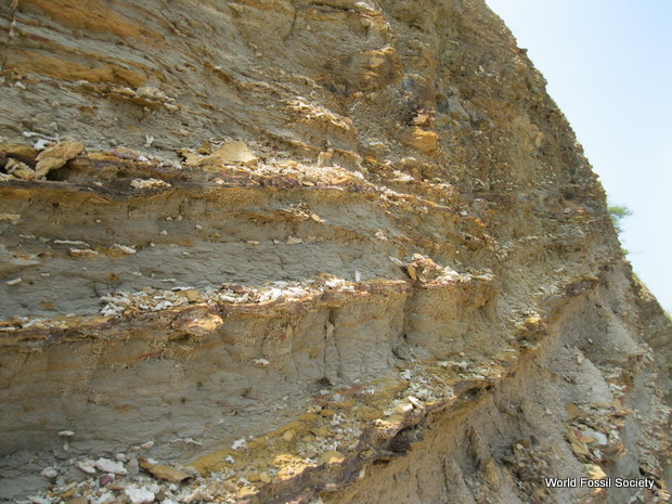
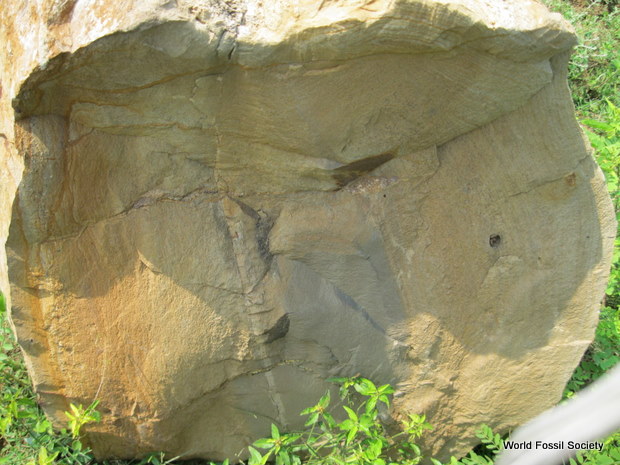
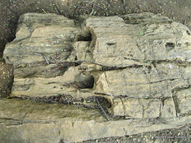
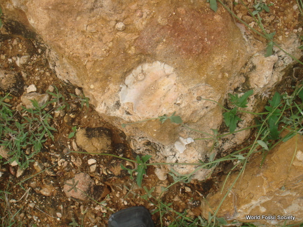
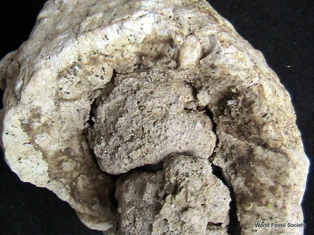
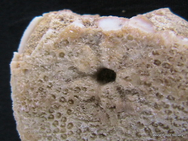
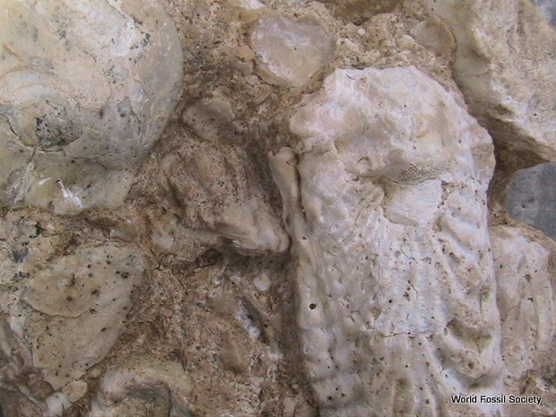
The Gogo Formation, a sedimentary rock formation in north-western Australia, has long been famous for yielding exquisitely preserved fossil fish. Among other things it contains placoderms, an extinct group that includes some of the earliest jawed fish.

A few years ago, an Australian research team work led by Prof. Trinajstic made the remarkable discovery that these fossils also contained soft tissues including nerve and muscle cells. Now they have collaborated with the research group of Professor Per Ahlberg, Uppsala University, and with the European Synchrotron (ESRF) in Grenoble, France, to document and reconstruct the musculature of the placoderms. “High contrast X-ray images were produced thanks to a powerful beam and a protocol developed for fossil imaging at the ESRF. This is unique in the world and has enabled us to “reconstruct” some fossilised muscles and document the muscles of neck and abdomen in these early jawed fish, without damaging or affecting the fossilised remains,” says Sophie Sanchez, one of the authors, from the ESRF and Uppsala University.

This is an alternative version with a pircture depicting the location in the fossil fish -- virtual thin section made in a nodule showing preserved bundles of muscles attached to the skull plate of a placoderm (fossil armored fish). (Credit: ESRF/Sophie Sanchez)

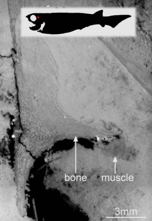
This is an alternative version with a pircture depicting the location in the fossil fish — virtual thin section made in a nodule showing preserved bundles of muscles attached to the skull plate of a placoderm (fossil armored fish). (Credit: ESRF/Sophie Sanchez)

These early vertebrates prove to have a well-developed neck musculature as well as powerful abdominal muscles — not unlike some human equivalents displayed on the beaches of the world every summer. Living fish, by contrast, usually have a rather simple body musculature without such specialisations.

“This shows that vertebrates developed a sophisticated musculature much earlier than we had thought” says Per Ahlberg, co-author of the project. “It also cautions against thinking that we can interpret fossil organisms simply by metaphorically draping their skeletons in the soft tissues of living relatives.”

High Diversity of Flying Reptiles in England 110 Million Years Ago

Brazilian paleontologists Taissa Rodrigues, of the Federal University of Espirito Santo, and Alexander W. A. Kellner, of the National Museum of the Federal University of Rio de Janeiro, have just presented the most extensive review yet available of toothed pterosaurs from the Cretaceous of England. The study features detailed taxonomic information, diagnoses and photographs of 30 species and was published in the open access journal ZooKeys.

Pterosaurs from the Cretaceous of England were first described by British naturalists Richard Owen and Harry Seeley in the 19th century, when little was known about the diversity of the group, resulting in the description of dozens of species, all based on very fragmentary remains, represented mostly by the tips of the snouts of these animals. However, more recent findings of pterosaur fossils have challenged views on their diversity.

The holotypes of the species recognized as valid by the researchers, put to the same scale, illustrate how diverse this pterosaur fauna was. (Credit: Dr. Taissa Rodrigues & NHMUK PV 39412, NHMUK PV R 1822, NHMUK PV 39409 and NHMUK PV 43074 - Natural History Museum)

The holotypes of the species recognized as valid by the researchers, put to the same scale, illustrate how diverse this pterosaur fauna was. (Credit: Dr. Taissa Rodrigues & NHMUK PV 39412, NHMUK PV R 1822, NHMUK PV 39409 and NHMUK PV 43074 – Natural History Museum)

Results show that these pterosaurs had a remarkable diversity in their appearances. Some species had head crests of different sizes and shapes, while others had none. Most had large teeth at the tip of their snouts and were fish eaters, but others had smaller teeth, suggesting different feeding preferences. The paleontologists were able to identify fourteen different species, belonging to at least five different genera, showing a greater diversity than previously thought.

Most of these fossils were found in a deposit known as the Cambridge Greensand, located in the eastern part of the country. This unit, one of the most important for the study of flying reptiles, records a past marine environment where the bones that were already fossilized and buried, were eroded, exposed to weathering, and then buried again. Cycles of erosion and burial must have taken place during several years. Due to this peculiarity, the pterosaur assemblage from this deposit probably presents temporal mixing of faunas, thus explaining the high diversity found.

Another find was that these English flying reptiles turned out to be closely related to species unearthed in northeastern Brazil and eastern China. According to Dr. Rodrigues, ‘This is very interesting, especially because the continents had already drifted apart. If these animals were migratory, we would expect to find the same species in all these deposits.’ Instead, the scientists have discovered that England, Brazil and China all had their own species and genera.

Analysis of fossils from other continents showed that this group of pterosaurs was already widespread in the whole planet 110 million years ago, and must have been important faunistic elements at this time of the Cretaceous period, being early bird competitors, before they went extinct a few million years later.

The flying reptiles


Rhamphorhynchus
A fossil of Rhamphorhynchus, an early pterosaur.


Ranging from the size of a sparrow to the size of an airplane, the pterosaurs (Greek for “wing lizards”) ruled the skies in the Jurassic and Cretaceous, and included the largest vertebrate ever known to fly: the late Cretaceous Quetzalcoatlus. The appearance of flight in pterosaurs was separate from the evolution of flight in birds and bats; pterosaurs are not closely related to either birds or bats, and thus provide a classic example of convergent evolution.


It was once thought that pterosaurs were not well adapted for active flight and relied largely on gliding and on the wind to stay in the air. However, based on analyses of pterosaur skeletal features (including work done by Berkeley’s own Kevin Padian), it is now thought that all but the largest pterosaurs could sustain powered flight. Pterosaurs had hollow bones, large brains with well-developed optic lobes, and several crests on their bones to which flight muscles attached. All of this is consistent with powered flapping flight.


Various pterosaurs


The largest pterosaur (Quetzalcoatlus, wonderfully named for the Aztec winged serpent god) had a wing span from eleven to twelve meters long (about forty feet). The wing’s main support was an amazingly elongated fourth digit in the hand. Fibers in the wing membrane added structural support and stiffness. At least some pterosaurs may have had some sort of hair-like body covering, which could very well mean that they were endothermic. Pterosaurs had a diverse range of head types, as you can tell from the pictures below. Their ability to fly probably allowed them to evolve into many niches, taking advantage of many different food sources, which would explain the range of skull morphology seen.


Pterosaurs consist of two main types (they do form a single (monophyletic) group, though): the “rhamphorhynchoids,” more properly termed the basal Pterosauria, which had long tails, and their descendants the “pterodactyloids,” which had shorter tails. Why is the term “rhamphorhynchoid” an invalid one? Since the later Pterosauria (the”pterodactyloids”) are the descendants of the basal Pterosauria, “rhamphorhynchoid” is a paraphyletic term, which phylogenetic researchers shy away from using. The basal Pterosauria (including Rhamphorhynchus, pictured at the top of this page) first appeared in the Late Triassic and all went extinct at the end of the Jurassic. The more derived pterosaurs (including Pteranodon, below) that were the descendants of this group appear first in Late Jurassic rocks, and the last of them died out at the end of the Cretaceous. Below is a mounted skeleton of Pteranodon ingens on display at the UCMP. Click on the picture to view an enlargement.


What was Pteranodon like?


Pteranodon skeleton
Pteranodon. Photo by Dave Smith, © 2005 UCMP.


The genus Pteranodon includes several species of large pterosaurs from the Cretaceous period in North America. As you can tell from this photo, it had a large crested head, a huge wingspan (some 20-25 feet; the UCMP specimen is about 22 feet), and a comparatively small body. This is deceiving; it looks like the head and wing bones were too bulky, and the hindlimbs appear small and weak. Not so; the bones of Pteranodon are actually completely hollow (about 1 millimeter thick!), and were quite light. The whole animal probably weighed about 25 pounds, only slightly heavier than the largest flying birds. The hindlimbs are actually perfectly sized for the body; Pteranodon would have been capable of bipedal terrestrial movement (but was no rapid runner, unlike its ancestors, some of whom seem to have been fast bipedal runners). The wing bones look thick because a large bone diameter is more vital for resisting the bending stresses involved in flight (as opposed to large bone thickness, which is important for resisting compressive forces, such as those imposed by the weight of a large body), so actually the wings of Pteranodon were more than adequate for flight.


Pteranodon was almost certainly a soaring animal; it used rising warm air to maintain altitude; a common strategy among large winged animals (among birds, albatrosses and vultures are adept at soaring). Its scooplike beak was used for snapping up fish as it soared over the oceans that it nested by. A good modern analog for Pteranodon would be the pelican.

Source: University of California Museum of Paleontology informations.

WFS Profiles: Edward Drinker Cope


Cope Edward Drinker 1840-1897

Cope Edward Drinker 1840-1897

Cope Edward Drinker 1840-1897

Cope Edward Drinker 1840-1897


Edward Drinker Cope was an American paleontologist and evolutionist. He was one of the founders of the Neo- Lamarckian school of evolutionary thought. This school believed that changes in developmental (embryonic) timing, not natural selection, was the driving force of evolution. In 1867, Cope suggested that most changes in species occured by coordinated additions to the ontogeny of all the individuals in a species. Speciation proceeded by the addition of stages to the end of embryonic sequences of development and by compression of earlier stages into the earlier parts of the developmental sequence. That is, a new developmental stage would be tacked onto the end of the developmental process, pushing the old end stage further back in development. Cope thought that groups of species that shared similar developmental patterns could be grouped into more inclusive groups (i.e. genera, families, and so on).

Cope tied together his notion of “accelerated growth” with Lamarckian ideas. He thought that parts of the body most in use would be most likely to become better developed at the expense of other, less used parts. That is, new ontogenetic additions would cause some body parts to become very well developed if those body parts were in heavy use. Divergence and diversity were created through changes in timing of development in different organ systems due to use. The more distantly related two species are, the more their developmental patterns will vary deep into ontogeny.

Cope led many natural history surveys in the American West for the precursors of the U.S. Geological Survey. He made many important finds on his trips, including dinosaur discoveries in western North America. He was primarily a herpetologist and mammalogist and he described many genera and species that are still used today.

Earthquake Acoustics Can Indicate If a Massive Tsunami Is Imminent

Stanford scientists have identified key acoustic characteristics of the 2011 Japan earthquake that indicated it would cause a large tsunami. The technique could be applied worldwide to create an early warning system for massive tsunamis.

On March 11, 2011, a magnitude 9.0 undersea earthquake occurred 43 miles off the shore of Japan. The earthquake generated an unexpectedly massive tsunami that washed over eastern Japan roughly 30 minutes later, killing more than 15,800 people and injuring more than 6,100. More than 2,600 people are still unaccounted for.

Now, computer simulations by Stanford scientists reveal that sound waves in the ocean produced by the earthquake probably reached land tens of minutes before the tsunami. If correctly interpreted, they could have offered a warning that a large tsunami was on the way.

Stanford scientists have identified key acoustic characteristics of the 2011 Japan earthquake that predicted it would cause a large tsunami. The technique could be applied worldwide to create an early warning system for tsunamis. See full video at: http://www.youtube.com/watch?v=4YxKSQvqgy8 (Credit: Courtesy of Stanford University / Video by Kurt Hickman)

Stanford scientists have identified key acoustic characteristics of the 2011 Japan earthquake that predicted it would cause a large tsunami. The technique could be applied worldwide to create an early warning system for tsunamis. See full video at: http://www.youtube.com/watch?v=4YxKSQvqgy8 (Credit: Courtesy of Stanford University / Video by Kurt Hickman)

Although various systems can detect undersea earthquakes, they can’t reliably tell which will form a tsunami, or predict the size of the wave. There are ocean-based devices that can sense an oncoming tsunami, but they typically provide only a few minutes of advance warning.

Because the sound from a seismic event will reach land well before the water itself, the researchers suggest that identifying the specific acoustic signature of tsunami-generating earthquakes could lead to a faster-acting warning system for massive tsunamis.

Discovering the signal

The finding was something of a surprise. The earthquake’s epicenter had been traced to the underwater Japan Trench, a subduction zone about 40 miles east of Tohoku, the northeastern region of Japan’s larger island. Based on existing knowledge of earthquakes in this area, seismologists puzzled over why the earthquake rupture propagated from the underground fault all the way up to the seafloor, creating a massive upward thrust that resulted in the tsunami.

Direct observations of the fault were scarce, so Eric Dunham, an assistant professor of geophysics in the School of Earth Sciences, and Jeremy Kozdon, a postdoctoral researcher working with Dunham, began using the cluster of supercomputers at Stanford’s Center for Computational Earth and Environmental Science (CEES) to simulate how the tremors moved through the crust and ocean.

The researchers built a high-resolution model that incorporated the known geologic features of the Japan Trench and used CEES simulations to identify possible earthquake rupture histories compatible with the available data.

Retroactively, the models accurately predicted the seafloor uplift seen in the earthquake, which is directly related to tsunami wave heights, and also simulated sound waves that propagated within the ocean.

In addition to valuable insight into the seismic events as they likely occurred during the 2011 earthquake, the researchers identified the specific fault conditions necessary for ruptures to reach the seafloor and create large tsunamis.

The model also generated acoustic data; an interesting revelation of the simulation was that tsunamigenic surface-breaking ruptures, like the 2011 earthquake, produce higher amplitude ocean acoustic waves than those that do not.

The model showed how those sound waves would have traveled through the water and indicated that they reached shore 15 to 20 minutes before the tsunami.

“We’ve found that there’s a strong correlation between the amplitude of the sound waves and the tsunami wave heights,” Dunham said. “Sound waves propagate through water 10 times faster than the tsunami waves, so we can have knowledge of what’s happening a hundred miles offshore within minutes of an earthquake occurring. We could know whether a tsunami is coming, how large it will be and when it will arrive.”

Worldwide application

The team’s model could apply to tsunami-forming fault zones around the world, though the characteristics of telltale acoustic signature might vary depending on the geology of the local environment. The crustal composition and orientation of faults off the coasts of Japan, Alaska, the Pacific Northwest and Chile differ greatly.

“The ideal situation would be to analyze lots of measurements from major events and eventually be able to say, ‘this is the signal’,” said Kozdon, who is now an assistant professor of applied mathematics at the Naval Postgraduate School. “Fortunately, these catastrophic earthquakes don’t happen frequently, but we can input these site specific characteristics into computer models — such as those made possible with the CEES cluster — in the hopes of identifying acoustic signatures that indicates whether or not an earthquake has generated a large tsunami.”

Dunham and Kozdon pointed out that identifying a tsunami signature doesn’t complete the warning system. Underwater microphones called hydrophones would need to be deployed on the seafloor or on buoys to detect the signal, which would then need to be analyzed to confirm a threat, both of which could be costly. Policymakers would also need to work with scientists to settle on the degree of certainty needed before pulling the alarm.

If these points can be worked out, though, the technique could help provide precious minutes for an evacuation.

The study is detailed in the current issue of the journal The Bulletin of the Seismological Society of America.

Secondary Cartilage Revealed in a Non-Avian Dinosaur Embryo

The skull and jaws of extant birds possess secondary cartilage, a tissue that arises after bone formation during embryonic development at articulations, ligamentous and muscular insertions. Using histological analysis, we discovered secondary cartilage in a non-avian dinosaur embryo, Hypacrosaurus stebingeri (Ornithischia, Lambeosaurinae). This finding extends our previous report of secondary cartilage in post-hatching specimens of the same dinosaur species. It provides the first information on the ontogeny of avian and dinosaurian secondary cartilages, and further stresses their developmental similarities. Secondary cartilage was found in an embryonic dentary within a tooth socket where it is hypothesized to have arisen due to mechanical stresses generated during tooth formation. Two patterns were discerned: secondary cartilage is more restricted in location in this Hypacrosaurus embryo, than it is in Hypacrosaurus post-hatchlings; secondary cartilage occurs at far more sites in bird embryos and nestlings than in Hypacrosaurus. This suggests an increase in the number of sites of secondary cartilage during the evolution of birds. We hypothesize that secondary cartilage provided advantages in the fine manipulation of food and was selected over other types of tissues/articulations during the evolution of the highly specialized avian beak from the jaws of their dinosaurian ancestors.

Secondary chondrogenesis investigated in hadrosaurid embryos.  (A) Reconstruction of the embryonic skull of Hypacrosaurus stebingeri, reproduced with permission [21] with anatomical locations 1, 2 and 3 in green. (B) Transverse section of the surangular of a Hadrosauridae indet. (MOR 1038). (C) Close-up of the red box in (B). The dorso-caudal face (Location 1) does not show any remnant of SC. (D) Coronal section of the maxilla of Hypacrosaurus stebingeri (MOR 559). (E) Close-up of the red box in (D). The bucco-caudal face of the maxilla (Location 2) does not show any remnants of SC. (F) Coronal section of the dentary of Hypacrosaurus stebingeri (MOR 559). (G) Close-up of the red box in (F). The arrow indicates a remnant of dentine. (H) Close-up of the red box in (G). (F) and (G) show alveolar bone (white asterisks) and incomplete alveoli with missing teeth (black asterisk; Location 3). (G) and (H) show a SC islet. All sections are shown under natural light. do, dorsal; la, labial; li, lingual; ro, rostral. doi:10.1371/journal.pone.0056937.g001

Secondary chondrogenesis investigated in hadrosaurid embryos.
(A) Reconstruction of the embryonic skull of Hypacrosaurus stebingeri, reproduced with permission [21] with anatomical locations 1, 2 and 3 in green. (B) Transverse section of the surangular of a Hadrosauridae indet. (MOR 1038). (C) Close-up of the red box in (B). The dorso-caudal face (Location 1) does not show any remnant of SC. (D) Coronal section of the maxilla of Hypacrosaurus stebingeri (MOR 559). (E) Close-up of the red box in (D). The bucco-caudal face of the maxilla (Location 2) does not show any remnants of SC. (F) Coronal section of the dentary of Hypacrosaurus stebingeri (MOR 559). (G) Close-up of the red box in (F). The arrow indicates a remnant of dentine. (H) Close-up of the red box in (G). (F) and (G) show alveolar bone (white asterisks) and incomplete alveoli with missing teeth (black asterisk; Location 3). (G) and (H) show a SC islet. All sections are shown under natural light. do, dorsal; la, labial; li, lingual; ro, rostral.
doi:10.1371/journal.pone.0056937.g001

Citation: Bailleul AM, Hall BK, Horner JR (2013) Secondary Cartilage Revealed in a Non-Avian Dinosaur Embryo. PLoS ONE 8(2): e56937. doi:10.1371/journal.pone.0056937

Editor: Peter Dodson, University of Pennsylvania, United States Of Amerca

How the turtles got their shells: Clues from fossils !

Through careful study of an ancient ancestor of modern turtles, researchers now have a clearer picture of how the turtles’ most unusual shell came to be. The findings, reported on May 30 in Current Biology, a Cell Press publication, help to fill a 30- to 55-million-year gap in the turtle fossil record through study of an extinct South African reptile known as Eunotosaurus.

The skeleton of the South African reptile Eunotosaurus africanus fills a gap in the early evolution of turtles and their enigmatic shell. (Credit: Tyler Lyson)

The skeleton of the South African reptile Eunotosaurus africanus fills a gap in the early evolution of turtles and their enigmatic shell. (Credit: Tyler Lyson)

“The turtle shell is a complex structure whose initial transformations started over 260 million years ago in the Permian period,” says Tyler Lyson of Yale University and the Smithsonian. “Like other complex structures, the shell evolved over millions of years and was gradually modified into its present-day shape.”

The turtle shell isn’t really just one thing — it is made up of approximately 50 bones. Turtles are the only animals that form a shell through the fusion of ribs and vertebrae. In all other animals, shells are formed from bony scales on the surface; they don’t stick their bones on the outsides of their bodies.

“The reason, I think, that more animals don’t form a shell via the broadening and eventually suturing together of the ribs is that the ribs of mammals and lizards are used to help ventilate the lungs,” Lyson says. “If you incorporate your ribs into a protective shell, then you have to find a new way to breathe!” Turtles have done just that, with the help of a muscular sling.

Until recently, the oldest known fossil turtles, dating back about 215 million years, had fully developed shells, making it hard to see the sequence of evolutionary events that produced them. That changed in 2008 with the discovery of Chinese Odontochelys semitestacea, a reptile about 220 million years old, which had a fully developed plastron — the belly side of the shell — but only a partial carapace on its back.

Eunotosaurus takes the turtle and its shell back another 40 million years or so. It had nine broadened ribs found only in turtles. And like turtles, it lacked the intercostal muscles running between its ribs. But Eunotosaurus didn’t have other features common to Odontochelys and turtles, including broad spines on their vertebrae.

Lyson says he and his colleagues now plan to investigate various other aspects of turtles’ respiratory systems, which allow them to manage with their ribs locked up into a protective outer shell. “It is clear that this novel lung ventilation mechanism evolved in tandem with the origin of the turtle shell,” he says.

Sturgeon Fish Are Evolutionary Speedsters

Efforts to restore sturgeon in the Great Lakes region have received a lot of attention in recent years, and many of the news stories note that the prehistoric-looking fish are “living fossils” virtually unchanged for millions of years.

But a new study by University of Michigan researchers and their colleagues reveals that in at least one measure of evolutionary change — changes in body size over time — sturgeon have been one of the fastest-evolving fish on the planet.

A lake sturgeon from the Great Lakes. (Credit: Photo courtesy of Michigan Sea Grant)

A lake sturgeon from the Great Lakes. (Credit: Photo courtesy of Michigan Sea Grant)

“Sturgeon are thought of as a living fossil group that has undergone relatively slow rates of anatomical change over time. But that’s simply not true,” said Daniel Rabosky, assistant professor in the U-M Department of Ecology and Evolutionary Biology and a curator of herpetology at the Museum of Zoology.

“Our study shows that sturgeon are evolving very quickly in some ways. They have evolved a huge range of body sizes. There are dwarf sturgeon the size of a bass and several other species that are nearly as big as a Volkswagen.”

The sturgeon finding is just one result in a wide-ranging study of the rates of species formation and anatomical change in fish. The work involved assembling one of the largest evolutionary trees ever created for any group of animals. The evolutionary relationships between nearly 8,000 species of fish are delineated in the branches of the tree, allowing the researchers to make inferences about all 30,000 or so species of ray-finned fish.

The study’s findings are scheduled for online publication in Nature Communications on June 6. Rabosky and Michael Alfaro of the University of California, Los Angeles, are the lead authors. U-M computational evolutionary biologist Stephen Smith is a co-author.

The main goal of the project was to test a longstanding idea in evolutionary biology that has anecdotal support but which had never been rigorously evaluated, Rabosky said. It was Charles Darwin who coined the term “living fossil” to describe extant creatures, such as the gar (another Great Lakes resident) and the lungfish, which have been present for many millions of years in the fossil record yet appear to have undergone very little anatomical change.

Paleontologists have long suspected that these observations reflect a fundamental coupling between the rates of species formation and anatomical change: groups of organisms that contain lots of species also seem to have greater amounts of anatomical variation, while groups with only a few species, such as the gar, lack much morphological variety.

Rabosky and his colleagues assembled a time-calibrated evolutionary tree for 7,864 living fish species using DNA sequence data and body-size information from publicly available databases. Their data set was so large that they had to develop new computer programs from scratch to analyze it.

The new computer models and the vast amount of data enabled the team to study the correlation between how quickly new species form and how rapidly they evolve new body sizes on a scale that had not previously been possible.

They found a strong correlation between the rates of species diversification and body size evolution across the more than 30,000 living species of ray-finned fish, which comprise the majority of vertebrate biological diversity.

“We’re basically validating a lot of ideas that have been out there since Darwin, but which had never been tested at this scale due to lack of data and the limits of existing technologies,” Rabosky said.

Most of the fish groups fall into one of two categories. Fish like the gar form species very slowly and show little range in body size. Others, like the salmon family — which includes salmon, trout, whitefish and char — do both: they form species quickly and have a wide range of body sizes.

Sturgeon have been around more than 100 million years and today consist of 29 species worldwide, including the lake sturgeon found in the Great Lakes. They don’t fit the general pattern found by Rabosky’s team; there are few sturgeon species but a great variety in body size.

“In that sense, they’re kind of an outlier,” Rabosky said.

In addition to Rabosky, Alfaro and Smith, authors of the Nature Communications paper are Francesco Santini of the Universita di Torino in Italy, Jonathan Eastman of the University of Idaho, Brian Sidlauskas of Oregon State University and Jonathan Chang of UCLA.

The research was supported by the National Science Foundation and the Miller Institute for Basic Research at the University of California, Berkeley.

Discovery of Oldest-Known Fossil Primate Skeleton announced

A mouse-sized fossil from China has provided remarkable new insights into the origin of primates.Xijun Ni points out the key features in the fossil

At 55 million years old, it represents the earliest known member of this broad group of animals that includes humans.scientists have called the diminutive creature Archicebus, which roughly translates as “ancient monkey”.They tell Nature magazine that its skeleton helps explain the branching that occurred at the very base of the primate evolutionary tree.

The team puts Archicebus on the line leading to tarsiers, a collection of small arboreal animals now found exclusively in south-east Asia.But its great age and primitive features mean it sits right at the base of this lineage, and so Archicebus therefore has much to say also about the emergence of the tarsiers’ sister grouping – the anthropoids, the primates that include monkeys, apes and us.

And it would suggest the first of these anthropoids were, likewise, petite creatures scurrying through the tropical canopies that grew to cover the Earth shortly after the dinosaurs’ extinction.

Archicebus
Artist’s impression: Archicebus lived on a very warm “forest planet”

“We are all very curious about the ancestors of primates, including those of human beings,” said Dr Xijun Ni from the Chinese Academy of Sciences, Beijing, China.

“From this almost complete skeleton, we can conclude that our ancestors were a kind of very small animal. It was very active and agile; and it lived in the trees and fed on insects,” he told BBC News.

The Archicebus fossil is preserved across two slate slabs. Most of the animal’s key bones are recorded, including exquisite impressions of its rear limbs and feet.

The specimen was originally discovered in the Jingzhou area of Hubei Province some 10 years ago by a local farmer, but Prof Ni and colleagues have deliberately taken their time to describe the creature and assess its importance.

Their investigation has included sending the delicate slate pieces to Grenoble, France, to be imaged at the European Synchrotron Radiation Facility (ESRF).

This giant machine uses brilliant X-rays to try to discern details of the skeleton that remain hidden inside the rock.

“There’s no way you can prepare the fossil any better to see its features because you would end up with a powder; it’s extremely fragile,” explained the ESRF’s Dr Paul Tafforeau. “But when you scan it with synchrotron light, you can virtually extract the bones without touching them. This gives you access to the general anatomy and we can achieve very high resolution.”

Brilliant X-rays were used to more fully visualise the creature’s skeleton

The forensic study has allowed the scientists to build a picture of the type of animal Archicebus was in life, and to gauge its relationship to all other primates.

Many people may be taken aback by the animal’s small size – a body just 71mm in length and an estimated weight of about 20-30g. But Archicebus gives us a good idea of what the very first primates on Earth would have looked like.

They would have emerged in a pivotal period, said senior Nature editor Dr Henry Gee, when the Earth was gripped in a period of outstanding global warming.

“At this time, 55 million years ago, the Earth was a jungle planet,” he told BBC News. “The whole Earth was covered with tropical jungle – full of trees for little scampery things to climb up and down on. It was an ideal time for primates to be evolving.”

Particular features of Archicebus‘ skeleton suggest it would have used a leap-and-grasp motion as it traversed through these great forests.

Its small pointy teeth indicate that it ate insects. Certainly, its size would have meant it had a high metabolic rate, and preying on insects would have satisfied its calorific needs.

And the creature’s relatively large eye sockets suggest it had good vision for hunting, but the team says the evidence points towards Archicebus being a daytime operator, not a nocturnal animal.

One of the most significant observations is the shape of its heel bone. Far from being like a tarsier’s calcaneus, the bone is more reminiscent of what one would expect in an anthropoid.

Dr Chris Beard from the Carnegie Museum of Natural History in Pittsburgh, US, recalls: “The heel, and the foot in general, was one of the most shocking parts of the anatomy of this fossil when we first saw it; because, frankly, the foot of this fossil primate looks like a small monkey, specifically like a marmoset.”

It emphasised, he said, Archicebus‘ proximity to the split in the two lineages.

“What is means is that the common ancestor of tarsiers and anthropoids had some features that looked more like anthropoids than tarsiers. And I guess we shouldn’t be so surprised by this.”

Primate family tree
  • Archicebus is placed right at the base of the tarsiers, which today are found in southeast Asia
  • Tarsiers and anthropoids (monkeys, apes and humans) belong to a primate clade known as haplorhines (dry noses)
  • The strepsirhines (wet noses) are a more distant grouping that includes the famous lemurs of Madagascar
  • This clade also includes an extinct collection of primates that scientists refer to as the adapids
  • One of its members, “Ida” (Darwinius), was proposed back in 2009 to be at the base of the primate lineage
  • The wider scientific community rejected this interpretation and put the 47-million-year-old Ida in with the adapids

Source: Article by

Oldest Known Eucalyptus Macrofossils Are from South America

The evolutionary history of Eucalyptus and the eucalypts, the larger clade of seven genera including Eucalyptus that today have a natural distribution almost exclusively in Australasia, is poorly documented from the fossil record. Little physical evidence exists bearing on the ancient geographical distributions or morphologies of plants within the clade. Herein, we introduce fossil material of Eucalyptus from the early Eocene (ca. 51.9 Ma) Laguna del Hunco paleoflora of Chubut Province, Argentina; specimens include multiple leaves, infructescences, and dispersed capsules, several flower buds, and a single flower. Morphological similarities that relate the fossils to extant eucalypts include leaf shape, venation, and epidermal oil glands; infructescence structure; valvate capsulate fruits; and operculate flower buds. The presence of a staminophore scar on the fruits links them to Eucalyptus, and the presence of a transverse scar on the flower buds indicates a relationship to Eucalyptus subgenus Symphyomyrtus. Phylogenetic analyses of morphological data alone and combined with aligned sequence data from a prior study including 16 extant eucalypts, one outgroup, and a terminal representing the fossils indicate that the fossils are nested within Eucalyptus. These are the only illustrated Eucalyptus fossils that are definitively Eocene in age, and the only conclusively identified extant or fossil eucalypts naturally occurring outside of Australasia and adjacent Mindanao. Thus, these fossils indicate that the evolution of the eucalypt group is not constrained to a single region. Moreover, they strengthen the taxonomic connections between the Laguna del Hunco paleoflora and extant subtropical and tropical Australasia, one of the three major ecologic-geographic elements of the Laguna del Hunco paleoflora. The age and affinities of the fossils also indicate that Eucalyptus subgenus Symphyomyrtus is older than previously supposed. Paleoecological data indicate that the Patagonian Eucalyptus dominated volcanically disturbed areas adjacent to standing rainforest surrounding an Eocene caldera lake.

A–B. MPEF-Pb 2329, A. Overall view showing the linear to lanceolate shape, the acute and acuminate apex, and the acute and decurrent base. B. Detail of the venation pattern; note the intramarginal, secondary, and intersecondary veins. C. MPEF-Pb 3729, detail of lamina showing island oil glands and higher order venation pattern. D. E. bridgesiana R.T. Baker, BH 37791, detail of the higher order venation pattern. E. E. camaldulensis Dehnh, detail of the lamina showing the island oil glands. F. E. tereticornis Sm., PBP-1003, overall view of a cleared leaf; note the general morphology of the lamina and the venation pattern. Scale bars: A, F, 1 cm; B–C, 1 mm; D–E, 5 mm. doi:10.1371/journal.pone.0021084.g002

A–B. MPEF-Pb 2329, A. Overall view showing the linear to lanceolate shape, the acute and acuminate apex, and the acute and decurrent base. B. Detail of the venation pattern; note the intramarginal, secondary, and intersecondary veins. C. MPEF-Pb 3729, detail of lamina showing island oil glands and higher order venation pattern. D. E. bridgesiana R.T. Baker, BH 37791, detail of the higher order venation pattern. E. E. camaldulensis Dehnh, detail of the lamina showing the island oil glands. F. E. tereticornis Sm., PBP-1003, overall view of a cleared leaf; note the general morphology of the lamina and the venation pattern. Scale bars: A, F, 1 cm; B–C, 1 mm; D–E, 5 mm.
doi:10.1371/journal.pone.0021084.g002


A. MPEF-Pb 3735, overall view of flower bud showing corolline operculum and transverse scar left after loss of the calyx. B. MPEF-Pb 3733, flower bud with hemispherical hypanthium and corolline operculum of coherent petals (arrows show petal margins). C. MPEF-Pb 3734, calycine scar (arrow) denotes the separation between the hypanthium and corolline operculum. D. E. microcorys, BH 37596, flower buds and open flower; note the hypanthium, and corolline operculum of coherent petals with visible margins (arrow) on the flower buds. E. MPEF-Pb 3738, bisexual flower with single style and stigma and numerous stamens. Scale bars: A–C, E, 2 mm; D, 3 mm. doi:10.1371/journal.pone.0021084.g003

A. MPEF-Pb 3735, overall view of flower bud showing corolline operculum and transverse scar left after loss of the calyx. B. MPEF-Pb 3733, flower bud with hemispherical hypanthium and corolline operculum of coherent petals (arrows show petal margins). C. MPEF-Pb 3734, calycine scar (arrow) denotes the separation between the hypanthium and corolline operculum. D. E. microcorys, BH 37596, flower buds and open flower; note the hypanthium, and corolline operculum of coherent petals with visible margins (arrow) on the flower buds. E. MPEF-Pb 3738, bisexual flower with single style and stigma and numerous stamens. Scale bars: A–C, E, 2 mm; D, 3 mm.
doi:10.1371/journal.pone.0021084.g003


A. MPEF-Pb 3750, infructescence composed of two umbellasters. B. MPEF-Pb 981, capsules in unbellaster. C. MPEF-Pb 3740, infructescence showing the morphology and orientation of the fruit disks. D. MPEF-Pb 3739, infructescence composed of at least three fruits showing valves and disks. E–F. MPEF-Pb 2374. E. Top view of a capsule showing corolline operculum scar (c), staminophore scar (s), valves (v), disk (d), and hypanthium (h). F. Side view of capsule showing the same features. Scale bars: A, B, D, 3 mm; C, 2 mm; E–F, 1 mm. doi:10.1371/journal.pone.0021084.g004

A. MPEF-Pb 3750, infructescence composed of two umbellasters. B. MPEF-Pb 981, capsules in unbellaster. C. MPEF-Pb 3740, infructescence showing the morphology and orientation of the fruit disks. D. MPEF-Pb 3739, infructescence composed of at least three fruits showing valves and disks. E–F. MPEF-Pb 2374. E. Top view of a capsule showing corolline operculum scar (c), staminophore scar (s), valves (v), disk (d), and hypanthium (h). F. Side view of capsule showing the same features. Scale bars: A, B, D, 3 mm; C, 2 mm; E–F, 1 mm.
doi:10.1371/journal.pone.0021084.g004


Citation: Gandolfo MA, Hermsen EJ, Zamaloa MC, Nixon KC, González CC, et al. (2011) Oldest Known Eucalyptus Macrofossils Are from South America. PLoS ONE 6(6): e21084. doi:10.1371/journal.pone.0021084

Editor: Andrew Allen Farke, Raymond M. Alf Museum of Paleontology, United States of America