Carcass of a 50-million-year-old fossil of a lizard

Even for the lucky few creatures that are preserved in the fossil record, soft tissues such as skin and feathers typically disappear over time. But a newly developed technique has found a way to bring them back to life in some cases. Researchers have now used the approach to resurrect the teeth and recognize the carcass of a 50-million-year-old fossil of a lizard, long thought to be merely preserved remnants of skin shed from the reptile.

“This is incredibly uncharted territory,” says Gregory Erickson, a vertebrate paleontologist at Florida State University in Tallahassee. “This technique reveals that there’s literally more to fossils than meets the eye.”

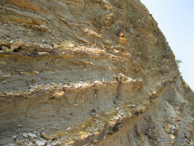
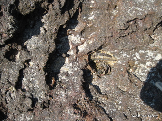
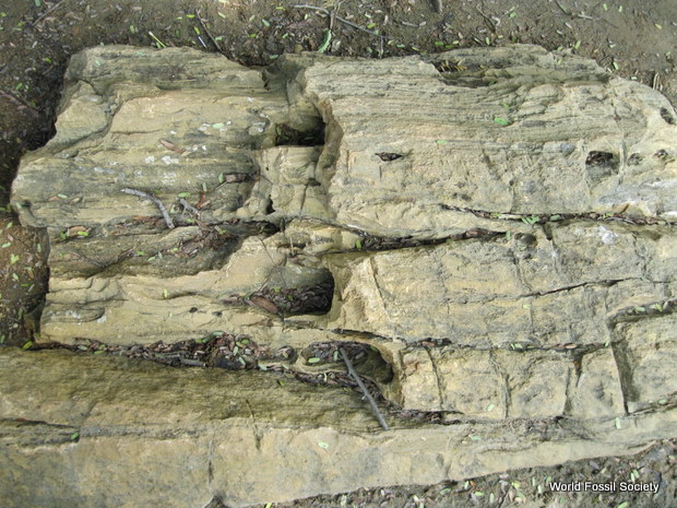
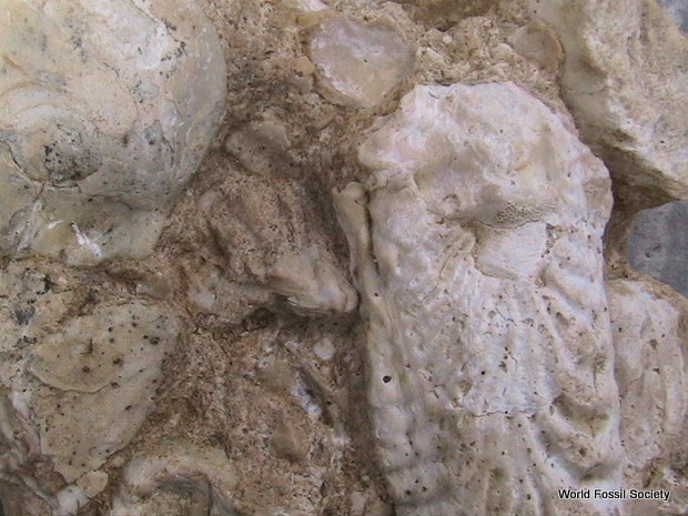
Discovered in the 1980s, the lizard fossil is one of only two known examples of reptile skin unearthed from the Green River Formation of the western United States, a finely layered mudstone best known for its exquisite fish fossils. Even though soft tissues are incredibly rare in the fossil record, being preserved only in unusual environmental circumstances, this lizard fossil survived the ages, says Phillip Manning, a vertebrate paleontologist at the University of Manchester in the United Kingdom. It’s easy to see the remnants of individual scales in the skin, but the rock doesn’t include any visible remains of bones or other hard tissue—a combination that led researchers to believe that the skin had been shed by a living creature and then preserved.

But recently, to learn more about the fossil, Manning and his colleagues turned to a relatively new x-ray analysis technique—dubbed synchrotron rapid scanning x-ray fluorescence—with surprising results. Instead of enabling scientists to see inside or through rock, he notes, the intense x-rays produced by this technique cause particular elements or compounds to fluoresce, revealing previously unrecognized chemical remnants that are invisible to the naked eye but persist in the rocks at very low concentrations.

When the researchers illuminated the fossil with x-rays that cause sulfur and copper to fluoresce, the skin remnants showed up in remarkable detail. But when they lit the fossil with x-rays that cause phosphorus to glow, the technique revealed many small spots in the lizard’s head where that element was concentrated—regularly spaced spots that appear where the creature’s jaws would have been. The arrangement prompted the researchers to interpret the traces of phosphorus as the chemical remnants of teeth. Because lizards don’t shed their teeth when they molt their skin, the technique reveals the unusual fossil to be the partially preserved remnants of a full carcass, the researchers report online this month in Applied Physics A: Materials Science & Processing.

X-ray vision. When researchers shone x-rays of certain wavelengths on what was thought to be merely a 50-million-year-old fossil of lizard skin (left), they discovered spots with high concentrations of phosphorus (dots, upper right) that they interpret as the chemical vestiges of teeth (teeth of separate jaws are outlined in blue and red, lower right). Credit: Edwards et al., Applied Physics A: Materials Science & Processing (2012)

X-ray vision. When researchers shone x-rays of certain wavelengths on what was thought to be merely a 50-million-year-old fossil of lizard skin (left), they discovered spots with high concentrations of phosphorus (dots, upper right) that they interpret as the chemical vestiges of teeth (teeth of separate jaws are outlined in blue and red, lower right).
Credit: Edwards et al., Applied Physics A: Materials Science & Processing (2012)

The fossil’s state of preservation reveals a lot about the environmental conditions where the carcass ended up, presumably after being washed into the lake soon after it died. Lake-bottom waters at this particular spot likely had little or no oxygen, enabling preservation of the skin. But the waters apparently were also acidic, which totally dissolved the creature’s bones and left only scant traces of its teeth. The chemical vestiges of the teeth were most likely preserved because tooth enamel typically has a low concentration of organic matter and large crystals of phosphate minerals, both of which render the teeth more resistant to decay.

The x-ray technique the team used “will open the curtain to a whole new way of studying extinct animals and the conditions in which they lived and died,” Manning says. Another benefit of the approach, he notes, is that it’s nondestructive.

Previous studies using the technique have revealed the chemical residues of pigments in feathers, providing insight into the color patterns that ancient birds might have sported. The technique also offers the opportunity to discern remnants of soft tissues that are only rarely preserved, such as the pigment-filled retinas of eyes, the ink sacs of ancient squid, and possibly other tissues such as muscles—at least as far as the naked eye is concerned.

Results of the new study “are fantastically interesting,” says Mark Norell, a vertebrate paleontologist at the American Museum of Natural History in New York City. “There’s a whole lot more preserved with fossils than we ever thought there was.”

Erickson agrees. “This technique will prompt paleontologists to revisit a lot of classic fossils,” he says. “Who knows what got missed during the first 150 years of paleontology?”

Internal Bone Structure Reveals Loading and Walking Behavior

The form and structure of bones change as a result of the forces to which they are subjected. Researcher Patrik Christen of Eindhoven University of Technology (TU/e) has used this fact to determine the load using the bone structure. This knowledge can be important in predicting the progress and treatment of bone diseases, as well as for understanding the walking behavior of extinct species. Christen will gain his PhD at TU/e on February 4.

An image of a micro-CT scan of the end of a human thigh bone. The cross-section shows the internal microstructure of the bone. (Credit: TU Eindhoven)

An image of a micro-CT scan of the end of a human thigh bone. The cross-section shows the internal microstructure of the bone. (Credit: TU Eindhoven)

The ability of the skeleton to adapt to the way in which it is loaded is well known as Wolff’s law. Bone tissue contains special cells that are sensitive to the mechanical forces acting on the bone. More bone mass is produced where the pressure on the bone is high, and bone tissue is lost where the pressure is low. This process is easily visible, for example in tennis players, where the heavily loaded playing arm gains extra bone mass, or in astronauts, where bone mass in the legs is lost after space travel as a result of the low load.

Algorithm Christen used the opposite of Wolff’s law, that the bone structure reveals information about the loads experienced in the past. He has developed an algorithm that searches micro-CT-scan images for load‑generated patterns in the microstructure of bones. The program translates these into a load distribution in the bone. Christen has used this method on wrist, hip and other bones, and his findings corresponded well to load data obtained in other studies. According to Christen his Orthopaedic Biomechanics research group at TU/e is the only one in the world that is able to reveal the load history of a bone on the basis of its microstructure.

Applications The method has a number of practical applications. For example knowledge of the bone loading patterns is important to allow better prediction of the development of bone diseases like osteoporosis, and to estimate the effectiveness of particular treatments. This can help to prevent painful and dangerous bone fractures. Christen’s method can also help to improve the shape of an implant to reduce the chance that it will later become detached. Finally, Christen expects that the method can be used to clarify the walking behavior of extinct species such as dinosaurs and our own ancestors, based on the bone structures found in fossils.

Patrik Christen will gain his PhD on Monday 4 February at Eindhoven University of Technology (TU/e) on his thesis entitled ‘Deciphering the Secret Message within Bone Microstructure’. His supervisor is prof.dr. Keita Ito, TU/e professor of Orthopaedic Biomechanics, and his Copromotor is dr.ir. Bert van Rietbergen.

First Artificial Enzyme Created by Evolution in a Test Tube

There’s a wobbly new biochemical structure in Burckhard Seelig’s lab at the University of Minnesota that may resemble what enzymes looked like billions of years ago, when life on earth began to evolve — long before they became ingredients for new and improved products, from detergents to foods and fuels.

Seelig created the fledgling enzyme by using directed evolution in the laboratory. Working with colleague Gianluigi Veglia, graduate student Fa-An Chao, and other team members, he subsequently determined its structure, which made its debut December 9 as an advance online publication in Nature Chemical Biology. Lab tests show that the enzyme (a type of RNA ligase, which connects two RNA molecules) functions like natural enzymes although its structure looks very different and it is flexible rather than rigid. Seelig speculates the new protein resembles primordial enzymes, before their current structures evolved.

3-D structure of the evolved enzyme (an RNA ligase), using 10 overlaid snapshots. In the top region, the overlays show the range of bending and folding flexibility in the amino acid chain that forms the molecule. The two gray balls are zinc ions. (Credit: University of Minnesota)

3-D structure of the evolved enzyme (an RNA ligase), using 10 overlaid snapshots. In the top region, the overlays show the range of bending and folding flexibility in the amino acid chain that forms the molecule. The two gray balls are zinc ions. (Credit: University of Minnesota)

Seelig and Veglia are professors in the College of Biological Sciences, where Fa-An Chao is a graduate student. Both faculty members have appointments in the Department of Biochemistry, Molecular Biology and Biophysics and Seelig is member of the BioTechnology Institute. Veglia also has an appointment in the Department of Chemistry in the university’s College of Science and Engineering.

While a handful of groups worldwide are developing artificial enzymes, they use rational design to construct the proteins on computers. Instead, the Seelig lab employs directed evolution. “To my knowledge, our enzyme is the only entirely artificial enzyme created in a test tube by simply following the principles of natural selection and evolution,” he says.

Rational enzyme design relies on preconceived notions of what a new enzyme should look like and how it should function. In contrast, directed evolution involves producing a large quantity of candidate proteins and screening several generations to produce one with the desired function. With this approach, the outcome isn’t limited by current knowledge of enzyme structure.

“Just as in nature, only the fittest survive after each successive generation,” Seelig explains. The process continues until it produces an enzyme that efficiently catalyzes a desired biochemical reaction. In this case, the new enzyme joins two pieces of RNA together.

“It’s kind of like giving typewriters to monkeys,” he says. “One monkey and one typewriter won’t produce anything clever. But if you have enough monkeys and typewriters, eventually one of them will write ‘to be or not to be’.” The lottery provides another analogy. “If you buy more tickets, you’re more likely to win,” Seelig says.

Like all proteins, the new RNA ligase enzyme is a chain of amino acids folded into a 3D structure, but the resemblance stops there. Natural enzymes, like all proteins, are made from alpha helices and beta strands. Seelig’s artificial enzyme lacks those structures. Instead, it forms around two metal ions and is not rigid. “Compared to enzymes we know from nature, the artificial enzyme has a rather unusual structure and dynamics,” Seelig says.

For decades, naturally occurring enzymes have been tweaked by industry to make industrial processes and products more effective. The ability to create enzymes from scratch using a natural process opens the door to a vast array of new products that provide business opportunities and improve quality of life without harmful environmental effects.

Going forward, Seelig plans to create enzymes with useful applications while he continues to explore the underlying basic science of enzyme structure and function, aiming to learn more about the origin of enzymes and how proteins evolve.

“Enzymes have always fascinated me,” he says. “It’s rewarding to do work that has practical applications yet provides the opportunity to better understand how life on earth evolved.”

Ups and Downs of Biodiversity After Mass Extinction

The climate after the largest mass extinction so far 252 million years ago was cool, later very warm and then cool again. Thanks to the cooler temperatures, the diversity of marine fauna ballooned, as paleontologists from the University of Zurich have reconstructed. The warmer climate, coupled with a high CO2 level in the atmosphere, initially gave rise to new, short-lived species. In the longer term, however, this climate change had an adverse effect on biodiversity and caused species to become extinct.

Amonoids peaked earlier after the vast mass extinction. (Credit: Image courtesy of University of Zurich)

Amonoids peaked earlier after the vast mass extinction. (Credit: Image courtesy of University of Zurich)

Until now, it was always assumed that it took flora and fauna a long time to recover from the vast mass extinction at the end of the Permian geological period 252 million years ago. According to the scientific consensus, complex ecological communities only began to reappear in the Middle Triassic, so 247 million years ago. Now, however, a Swiss team headed by paleontologist Hugo Bucher from the University of Zurich reveals that marine animal groups such as ammonoids and conodonts (microfossils) already peaked three or four million years earlier, namely still during the Early Triassic.

The scientists chart the temperature curves in detail in Nature Geoscience, demonstrating that the climate and the carbon dioxide level in the atmosphere fluctuated greatly during the Early Triassic and what impact this had on marine biodiversity and terrestrial plants.

Alternate cooler and very warm phases

For their climate reconstruction, Bucher and his colleagues analyzed the composition of the oxygen isotopes in conodonts, the remains of chordates that once lived in the sea. According to the study, the climate at the beginning of the Triassic 249 million years ago was cool. This cooler phase was followed by a brief very warm climate phase. At the end of the Early Triassic, namely between 247.9 and 245.9 million years ago, cooler conditions resumed.

Climate and carbon cycle influence biodiversity

The scientists then examined the impact of the climate on the development of flora and fauna. “Biodiversity increased most in the cooler phases,” explains paleontologist Bucher. “The subsequent extremely warm phase, however, led to great changes in the marine fauna and a major ecological shift in the flora.” Bucher and his team can reveal that this decline in biodiversity in the warm phases correlates with strong fluctuations in the carbon isotope composition of the atmosphere. These, in turn, were directly related to carbon dioxide gases, which stemmed from volcanic eruptions in the Siberian Large Igneous Province.

Species emerge and die out

Through the climatic changes, conodont and ammonoid faunae were initially able to recover very quickly during the Early Triassic as unusually short-lived species emerged. However, the removal of excess CO2 by primary producers such as algae and terrestrial plants had adverse effects in the long run: The removal of these vast amounts of organic matter used up the majority of the oxygen in the water. Due to the lack of oxygen in the oceans, many marine species died out. “Our studies reveal that greater climatic changes can lead to both the emergence and extinction of species. Thus, it is important to consider both extinction rates and the rate at which new species emerged,” says Bucher.

Bucher and his colleagues are convinced that climate changes and the emission of volcanic gases were key drivers of biotic recovery in the oceans during the Early Triassic: Cooler climate phases encourage biological diversification. Warmer climate phases and very high CO2 levels in the atmosphere, however, can have a harmful impact on biodiversity.

Poisonous prehistoric ‘raptor’ discovered by research team from Kansas and China

A group of University of Kansas researchers working with Chinese colleagues have discovered a venomous, birdlike raptor that thrived some 128 million years ago in China. This is the first report of venom in the lineage that leads to modern birds.

“This thing is a venomous bird for all intents and purposes,” said Larry Martin, KU professor and curator of vertebrate paleontology at the Natural History Museum and Biodiversity Institute. “It was a real shock to us and we made a special trip to China to work on this.”

The KU-China team’s findings will be published in the early edition of the Proceedings of the National Academy of Sciences during the week of Dec. 21.

“We think it’s going to make a big splash,” said Martin.

The article’s authors are Enpu Gong, geology department at Northeastern University in Shenyang, China, and researchers Martin, David Burnham and Amanda Falk at the KU Natural History Museum and Biodiversity Institute.

The dromaeosaur or raptor, Sinornithosaurus (Chinese-bird-lizard), is a close relative to Velociraptor. It lived in prehistoric forests of northeastern China that were filled with a diverse assemblage of animals including other primitive birds and dinosaurs.

“This is an animal about the size of a turkey,” said Martin. “It’s a specialized predator of small dinosaurs and birds. It was almost certainly feathered. It’s a very close relative of the four-winged glider called Microraptor.”

The venom most likely sent the victim into rapid shock, shrinking the odds of retaliation, escape or piracy from other predators while the raptor manipulated its prey.

“You wouldn’t have seen it coming,” said Burnham. “It would have swooped down behind you from a low-hanging tree branch and attacked from the back. It wanted to get its jaws around you. Once the teeth were embedded in your skin the venom could seep into the wound. The prey would rapidly go into shock, but it would still be living, and it might have seen itself being slowly devoured by this raptor.”

This image of fossilized Sinornithosaurus shows the raptor's long, grooved fangs. It lived in prehistoric forests of northeastern China that were filled with a diverse assemblage of animals including other primitive birds and dinosaurs. - David A. Burnham, PhD University of Kansas Biodiversity Institute     This image of fossilized Sinornithosaurus shows the raptor's long, grooved fangs. It lived in prehistoric forests of northeastern China that were filled with a diverse assemblage of animals including other primitive birds and dinosaurs. - David A. Burnham, PhD University of Kansas Biodiversity Institute

This image of fossilized Sinornithosaurus shows the raptor’s long, grooved fangs. It lived in prehistoric forests of northeastern China that were filled with a diverse assemblage of animals including other primitive birds and dinosaurs. – David A. Burnham, PhD University of Kansas Biodiversity Institute 

The genus had special depressions on the side of its face thought by the investigators to have housed a poison gland, connected by a long lateral depression above the tooth row that delivered venom to a series of long, grooved teeth on the upper jaw. This arrangement is similar to the venom-delivery system in modern rear-fanged snakes and lizards. The researchers believe it to be specialized for predation on birds.

“When we were looking at Sinornithosaurus, we realized that its teeth were unusual, and then we began to look at the whole structure of the teeth and jaw, and at that point, we realized it was similar to modern-day snakes,” Martin said.

Sinornithosaurus is represented by at least two species. These specimens have features consistent with a primitive venom-delivery system. The KU-China research team said it was a low-pressure system similar to the modern Beaded lizard, Heloderma, however the prehistoric Sinornithosaurus had longer teeth to break through layers of feathers on its bird victims.

The discovery of features thought to be associated with a venom-delivery system in Sinornithosaurus stemmed from a study of the anatomy and ecology of Microraptor by the joint Chinese-KU team. They now are seeking to discover if Microraptor may have possessed a similar poison-delivery system.

Note: This story has been adapted from a news release issued by the University of Kansas

Extinction Rates Not as Bad as Feared … for Now: Scientists Challenge Common Belief

Concerns that many animals are becoming extinct, before scientists even have time to identify them, are greatly overstated, according Griffith University researcher, Professor Nigel Stork. Professor Stork has taken part in an international study, the findings of which have been detailed in “Can we name Earth’s species before they go extinct?” published in the journal Science.

Deputy Head of the Griffith School of Environment, Professor Stork said a number of misconceptions have fueled these fears, and there is no evidence that extinction rates are as high as some have feared.

The rate of species extinction may not be as bad as first thought, but recording of species is still a mammoth task. (Credit: Griffith University)

“Surprisingly, few species have gone extinct, to our knowledge. Of course, there will have been some species which have disappeared without being recorded, but not many we think,” Professor Stork said.

Professor Stork said part of the problem is that there is an inflated sense of just how many animals exist and therefore how big the task to record them.

“Modern estimates of the number of eukaryotic species have ranged up to 100 million, but we have estimated that there are around 5 million species on the planet (plus or minus 3 million).”

And there are more scientists than ever working on the task. This contrary to a common belief that we are losing taxonomists, the scientists who identify species.

“While this is the case in the developed world where governments are reducing funding, in developing nations the number of taxonomists is actually on the rise.

“World-wide there are now two to three times as many taxonomist describing species as there were 20 years ago.”

Even so, Professor Stork says the scale of the global taxonomic challenge is not to be underestimated.

“The task of identifying and naming all existing species of animals is still daunting, as there is much work to be done.”

Other good news for the preservation of species is that conservation efforts in the past few years have done a good job in protecting some key areas of rich biodiversity.

But the reprieve may be short-lived.

“Climate change will dramatically change species survival rates, particularly when you factor in other drivers such as overhunting and habitat loss,” Professor Stork said.

“At this stage we have no way of knowing by how much extinction rates may escalate.

“But once global warming exceeds the 2 degree barrier, we can expect to see the scale of loss many people already believe is happening. Higher temperature rises coupled with other environmental impacts will lead to mass extinctions”

Prehistoric Ghosts Revealing New Details: Synchrotron Helps Identify Previously Unseen Anatomy Preserved in Fossils

Scientists at The University of Manchester have used synchrotron-based imaging techniques to identify previously unseen anatomy preserved in fossils.

Their work on a 50-million-year-old lizard skin identified the presence of teeth (invisible to visible light), demonstrating for the first time that this fossil animal was more than just a skin moult. This was only possible using some of the brightest light in the universe, X-rays generated by a synchrotron.

An illustration of how the lizard fossil was scanned using the X-ray process in the synchrotron. (Credit: Image courtesy of University of Manchester)

An illustration of how the lizard fossil was scanned using the X-ray process in the synchrotron. (Credit: Image courtesy of University of Manchester)

Dr Phil Manning, Dr Nick Edwards, Dr Roy Wogelius and colleagues from the Palaeontology Research group used Synchrotron Rapid Screening X-ray Fluorescence at the Stanford Synchrotron Radiation Lightsource in California to map the chemical make up of a rare fossil lizard skin. This cutting edge technology uses powerful x-rays that enabled the team to map the presence of phosphorus from teeth in this ancient reptile.

The relative position of the phosphorous in the skin fossil helped the scientists identify the type of lizard. They believe that the more elongated snout in conjunction with the general jaw shape bears a strong resemblance to a shinisaurid lizard (Bahndwivici ammoskius). The presence of phosphorous also demonstrates for the first time that the fossil skin is more than just a moult, as no lizards can shed their teeth along with their skin!

Talking about the images Dr Manning said: “Finding the presence of teeth changes almost everything we thought we knew about this fossil. We can identify the type of lizard for the first time, based upon the geometry of the teeth. Our findings also raise some fascinating questions about what happened to the animal after its death. What wiped out its bones but preserved the skin and the ghost of its teeth?”

The results of the analysis of the fossil using the synchrotron have been published in the journal Applied Physics A. It adds to the growing weight of evidence that powerful synchrotrons offer advanced fossil analysis.

Dr Manning also said: “The technique permits us to tease-out chemical information from fossils, information that you simply cannot see with the naked eye. Such chemical maps can help us see ‘ghosts’ of original biological structures that only remain in very dilute concentrations in the fossil.”

Dr Nick Edwards, a senior author on the paper, said: “This technology changes how we view taphonomy (the study of decaying organisms and how they are fossilized). We can now start looking for traces of animals that are totally invisible in visible light through analysing the bright chemical signature that appear under the powerful gaze of the synchrotron. This ‘x-ray vision’ will enable palaeontologists to add important information to the biology, anatomy and preservation of ancient life.”

The team worked with a leading geochemist, Dr Roy Wogelius (also a senior author), who was instrumental in the development of the techniques deployed by the Palaeontology Research Group. Dr Wogelius said: “These techniques are beginning to redefine the way we study Life on Earth. It is simply fascinating to work with biologists, physicists, chemists and palaeobiologists because at the crossroads of these disciplines lay many new discoveries for science.”

Dr Manning and his team hope to analyse more fossils using the Standford Synchrotron Radiation Lightsource and also the UK based Diamond Lightsource, to reveal new findings from ancient worlds by uncovering the subtle echoes of life left for science to find and interpret.

New Dinosaur Fossil Challenges Bird Flight Origins Theories

The discovery of a new bird-like dinosaur from the Jurassic period challenges widely accepted theories on the origin of flight.

Co-authored by Dr Gareth Dyke, Senior Lecturer in Vertebrate Palaeontology at the University of Southampton, the paper describes a new feathered dinosaur about 30 cm in length which pre-dates bird-like dinosaurs that birds were long thought to have evolved from.

Over many years, it has become accepted among palaeontologists that birds evolved from a group of dinosaurs called theropods from the Early Cretaceous period of Earth’s history, around 120-130 million years ago. Recent discoveries of feathered dinosaurs from the older Middle-Late Jurassic period have reinforced this theory.

Reconstruction of Eosinopteryx. (Credit: Royal Belgian Institute of Natural Sciences)

The new ‘bird-dinosaur’ Eosinopteryx described in Nature Communications this week provides additional evidence to this effect.

“This discovery sheds further doubt on the theory that the famous fossil Archaeopteryx — or “first bird” as it is sometimes referred to — was pivotal in the evolution of modern birds,” says Dr Dyke, who is based at the National Oceanography Centre, Southampton.

“Our findings suggest that the origin of flight was much more complex than previously thought.”

The fossilised remains found in north-eastern China indicate that, while feathered, this was a flightless dinosaur, because of its small wingspan and a bone structure that would have restricted its ability to flap its wings.

The dinosaur also had toes suited to walking along the ground and fewer feathers on its tail and lower legs, which would have made it easier to run.

Dr Gareth Dyke is also Programme Leader for a new one-year MRes in Vertebrate Palaeontology, which offers potential students the chance to study the evolution and anatomy of vertebrates, in order to inform and increase our understanding of the workings of modern day creatures.

Dr Dyke’s co-authors are Pascal Godefroit of the Royal Belgian Institute of Natural Sciences, Helena Demuynck of Earth System Science Vrije Universiteit Brussel, Dongyu Hu of Paleontological Institute Shenyang Normal University China and Key Laboratory of Vegetation Ecology Northeast Normal University China, François Escuillié of Eldonia France and Philippe Claeys of Jilin University Geological Museum China

Sex of Early Birds Suggests Dinosaur Reproductive Style

In a paper published in Nature Communications on January 22, 2013, a team of paleontologists including Dr. Luis Chiappe, Director of the Natural History Museum of Los Angeles County’s (NHM) Dinosaur Institute, has discovered a way to determine the sex of a prehistoric bird species.

Reconstruction of Confuciusornis sanctus. (Credit: Stephanie Abramowicz, NHM Dinosaur Institute)

Confuciusornis sanctus, a 125-million-year-old Mesozoic bird, had remarkable differences in plumage — some had long, almost body length ornamental tail feathers, others had none — features that have been interpreted as the earliest example of avian courtship. However, the idea that male Confuciusornis birds had ornamental plumage, and females did not, has not been proven until now. Chiappe and the team studied hundreds of Confuciusornis fossils unearthed from rocks deposited at the bottom of ancient lakes in what is today northeastern China and found undisputed evidence of the gender difference: medullary bone.

Chiappe conducted the study with Anusuya Chinsamy of the Department of Biological Sciences, University of Cape Town, South Africa; Jesús Marugán-Lobón of Madrid’s Universidad Autonóma, Cantoblanco; Gao Chunling and Zhang Fengjiao of the Dalian Natural History Museum in China.

“Our discovery provides the first case of sex identification in an ancient bird, an animal closely related to dinosaurs, such as the famous Velociraptor,” said Chiappe. “When people visit dinosaur exhibits, they often want to know if the skeletons are male or female. We have nicknames like Thomas and Sue, but of all the thousands of skeletons of dinosaurs or early birds found around the world, only the sex of a few has been determined.”

According to Chinsamy, the bone histologist on the team, “Just like modern hens, female Confuciusornis birds that lived 125 million years ago deposited this special bone inside their long bones, and then used it to make the calcium-rich eggshells.” Finding such tissue — present during a short period of time in reproductively active females — in a specimen that lacked long feathers proved that those birds without ornamental plumage are females.

“This now permits us to assess gender differences in growth and development of this Mesozoic bird,” she said.

But while this discovery offers evidence that both early and modern female avian species were essentially using the same physiological strategy to reproduce, it also spotlights an important difference in when they sexually matured.

“In human terms, knowing the sex of these specimens sheds light on when these early birds begin puberty,” said Chiappe, “Now we know that early birds began reproducing way before they were full grown, a pattern that contrasts with what we know of living birds, which typically begin reproducing after they reach full body size.” In that way, ancient birds produced offspring like dinosaurs, which also began to reproduce before they were fully grown.

The specimens, housed at the Dalian Natural History Museum in northeastern China, had been excavated from rocks formed at the bottom of ancient lakes in a forested environment surrounded by volcanoes. Ancient catastrophes, presumably related to volcanic eruptions, killed large numbers of birds and other animals, whose bodies were buried deep in the lake mud that helped minimize decay and preserving the organs, skeletons, and plumage. “This discovery is part of the big picture of understanding the early evolution of birds,” Chiappe said, “and how living birds became what they are.”

How dinosaurs measure up with laser imaging

Karl Bates and his colleagues in the palaeontology and biomechanics research group have reconstructed the bodies of five dinosaurs, two T. rex (Stan at the Manchester Museum and the Museum of the Rockies cast MOR555), an Acrocanthosaurus atokensis, a Strutiomimum sedens and an Edmontosaurus annectens.

The team, whose findings are published in the Public Library of Science journal PLoS ONE today (19th February 2009), found that the smaller Museum of the Rockies T. rex could have weighed anywhere between 5.5 and 7 tonnes, while the larger specimen (Stan) might have weighed as much as 8 tonnes.

Acrocanthosaurus atokensis was a large predatory dinosaur that looked like T. rex but with large spines on its back and roamed the earth much earlier in the mid Cretaceous period, around 110M years ago. The team suggest Acrocanthosaurus probably weighed in at a similar mass to MOR555 and other medium sized adult T. rex at about 6 tonnes.

The Strutiomimum sedens, whose name means “ostrich mimic”, lived alongside T. rex in the late Cretaceous period and probably weighed somewhere between 0.4 – 0.6 tonnes

The reconstruction of Edmontosaurus annectens, a plant-eating hadrosaur was based on a juvenile specimen, but still weighed in at between 0.8 – 0.95 tonnes. As adults, some hadrosaurs grew as big as T. rex, again living in the late Cretaceous period.

The team used laser scanning (LiDAR) and computer modelling methods to create a range of 3D models of the specimens, attempting to reconstruct their body sizes and shape as in life. The laser scanner images the full mounted skeleton, resulting in a detailed 3D model in which each bone retains its spatial position and articulation. This provides a high resolution skeletal framework around which the body cavity and internal organs such as stomach, lungs and air sacs can be reconstructed. This has allowed calculation of body segment masses, centres of mass and moments of inertia for each animal – all the information that is needed to analyse body movements.

Having created their ‘best-guess’ reconstruction of each animal, they then varied the volumes of body segments and respiratory organs to find the maximum plausible range of mass for the animals. Even scientists cannot be sure exactly how fat or thin animals like T. rex were in life, and the team were interested in exactly how broad the range of possible values were for body mass. They believe that the lower weight estimates are most likely to be correct as there is no good reason for the dinosaurs to weigh more than they need to as this would affect their speed, energy use and demands on the respiratory system.

The team also measured the body mass of an ostrich, as an existing subject that would show how accurate their technique was, and found the results to be correct.

They will now use the results to further investigate the locomotion of dinosaurs, specifically how they ran.

Karl said: “Our technique allows people to see and decide for themselves how fat or thin the dinosaurs might have been in life. You can see the skeleton with a belly. Anyone from a five-year-old to a Professor can see it and say, ‘I think this reconstruction is too fat or too thin’.

He added: “This study will help us in our research on how dinosaurs ran in 3-D rather than 2-D as in previous studies.

“Reconstructing more dinosaurs in such detail will allow us to examine changes in body mass and particularly centre of mass as they evolved. As we know, dinosaurs evolved into birds. As they did so, the centre of mass moved forward and different walking styles evolved. Although the dinosaurs we have reconstructed are not very close relatives of the birds, we can nevertheless see a small forwards movement in the position of the centre of mass from Acrocanthosaurus atokensis to the T. rex, which lies closer to modern birds on the evolutionary lines.”

Note: This story has been adapted from a news release issued by the University of Manchester