WFS News: Mongolian micro fossils point to the rise of animals on Earth.

@ WFS,World Fossil Society,Riffin T Sajeev,Russel T Sajeev

A Yale-led research team has discovered a cache of embryo-like microfossils in northern Mongolia that may shed light on questions about the long-ago shift from microbes to animals on Earth.

Called the Khesen Formation, the site is one of the most significant for early Earth fossils since the discovery of the Doushantuo Formation in southern China nearly 20 years ago. The Dousantuo Formation is 600 million years old; the Khesen Formation is younger, at about 540 million years old.

This is an image of assorted microfossils from the Ediacaran Khesen Formation, Mongolia. Each fossil is on the order of 200 microns maximum dimension. Credit: Yale University

This is an image of assorted microfossils from the Ediacaran Khesen Formation, Mongolia. Each fossil is on the order of 200 microns maximum dimension.Credit: Yale University

“Understanding how and when animals evolved has proved very difficult for paleontologists. The discovery of an exceptionally well-preserved fossil assemblage with animal embryo-like fossils gives us a new window onto a critical transition in life’s history,” said Yale graduate student Ross Anderson, first author of a study in the journal Geology.

The new cache of fossils represents eight genera and about 17 species, comprising tens to hundreds of individuals. Many of them are spiny microfossils called acritarchs, which are roughly 100 microns in size — about one-third the thickness of a fingernail.

The Khesen Formation is located to the west of Lake Khuvsgul in northern Mongolia. “This site was of particular interest to us because it had the right type of rocks — phosphorites — that had preserved similar organisms in China,” Anderson said.The discovery may help scientists confirm a much earlier date for the existence of Earth ecosystems with animals, rather than just microbes. For two decades, researchers have debated the findings at the Doushantuo Formation, with no resolution. If confirmed as animals, these microfossils would represent the oldest animals to be preserved in the geological record.

The other authors of the study are Derek Briggs, Yale’s G. Evelyn Hutchinson Professor of Geology and Geophysics and curator at the Yale Peabody Museum of Natural History; Sean McMahon, a postdoctoral fellow in the Briggs lab; Francis Macdonald of Harvard; and David Jones of Amherst College.The researchers said the Khesen Formation should provide scientists with additional information for years to come.

“This study is only the tip of the iceberg, as most of the fossils derive from only two samples,” Anderson said. Since the original discovery, the Yale team has worked with Harvard and the Mongolian University of Science and Technology to sample several additional sites within the formation.

  1. Ross P. Anderson, Francis A. Macdonald, David S. Jones, Sean McMahon, Derek E.G. Briggs. Doushantuo-type microfossils from latest Ediacaran phosphorites of northern Mongolia. Geology, 2017; DOI: 10.1130/G39576.1
  2. Yale University. “Mongolian microfossils point to the rise of animals on Earth.” ScienceDaily. ScienceDaily, 23 October 2017. <www.sciencedaily.com/releases/2017/10/171023123530.htm>.

 @ WFS,World Fossil Society,Riffin T Sajeev,Russel T Sajeev

WFS News: Scientists determine source of world’s largest mud eruption

@WFS,World Fossil Society,Riffin T Sajeev,Russel T Sajeev

On May 29, 2006, mud started erupting from several sites on the Indonesian island of Java. Boiling mud, water, rocks and gas poured from newly-created vents in the ground, burying entire towns and compelling many Indonesians to flee. By September 2006, the largest eruption site reached a peak, and enough mud gushed on the surface to fill 72 Olympic-sized swimming pools daily.

Indonesians frantically built levees to contain the mud and save the surrounding settlements and rice fields from being covered. The eruption, known as Lusi, is still ongoing and has become the most destructive ongoing mud eruption in history. The relentless sea of mud has buried some villages 40 meters (130 feet) deep and forced nearly 60,000 people from their homes. The volcano still periodically spurts jets of rocks and gas into the air like a geyser. It is now oozing around 80,000 cubic meters (3 million cubic feet) of mud each day — enough to fill 32 Olympic-sized pools. Watch a video of the Lusi eruption here: https://www.youtube.com/watch?v=1PXS1OIAD4o&feature=youtu.be

Now, more than 11 years after it first erupted, researchers may have figured out why the mudflows haven’t stopped: deep underground, Lusi is connected to a nearby volcanic system.

In a new study, researchers applied a technique geophysicists use to map Earth’s interior to image the area beneath Lusi. The images show the conduit supplying mud to Lusi is connected to the magma chambers of the nearby Arjuno-Welirang volcanic complex through a system of faults 6 kilometers (4 miles) below the surface.

On May 29, 2006, mud started erupting from several sites on the Indonesian island of Java and hasn’t stopped since. The eruption became known as Lusi and is the most destructive ongoing mud eruption in history. Credit: Adriano Mazzini/The Lusi Lab Project

On May 29, 2006, mud started erupting from several sites on the Indonesian island of Java and hasn’t stopped since. The eruption became known as Lusi and is the most destructive ongoing mud eruption in history.Credit: Adriano Mazzini/The Lusi Lab Project

Volcanoes can be connected to each other deep underground and scientists suspected Lusi and the Arjuno-Welirang volcanic complex were somehow linked, because previous research showed some of the gas Lusi expels is typically found in magma. But no one had yet shown that Lusi is physically connected to Arjuno-Welirang.

The researchers discovered that the scorching magma from the Arjuno-Welirang volcano has essentially been “baking” the organic-rich sediments underneath Lusi. This process builds pressure by generating gas that becomes trapped below the surface. In Lusi’s case, the pressure grew until an earthquake triggered it to erupt.

Studying the connection of these two systems could help scientists to better understand how volcanic systems evolve, whether they erupt magma, mud or hydrothermal fluids.

“We clearly show the evidence that the two systems are connected at depth,” said Adriano Mazzini, a geoscientist at CEED — University of Oslo and lead author of the new study in the Journal of Geophysical Research: Solid Earth, a journal of the American Geophysical Union. “What our new study shows is that the whole system was already existing there — everything was charged and ready to be triggered.”

Finding a connection

Java is part of a volcanic island arc, formed when one tectonic plate subducts below another. As the island rose upward out of the sea, volcanoes formed along its spine, with basins of shallow water between them. Lusi’s mud comes from sediments laid down in those basins while the island was still partially submerged underwater.

Mazzini has been studying Lusi since soon after the eruption began. Two years ago, the study’s authors installed a network of 31 seismometers around Lusi and the neighboring volcanic complex. Researchers typically use seismometers to measure ground shaking during earthquakes, but scientists can also use them to create three-dimensional images of the areas underneath volcanoes.

Using 10 months of data recorded by the seismometers, Mazzini and his colleagues imaged the area below Lusi and the surrounding volcanoes. The images showed a tunnel protruding from the northernmost of Arjuno-Welirang’s magma chambers into the sedimentary basin where Lusi is located. This allows magma and hydrothermal fluids originating in the mantle to intrude into Lusi’s sediments, which triggers massive reactions and creates gas that generates high pressure below Earth’s surface. Any perturbation — like an earthquake — can then trigger this system to erupt.

“It’s just a matter of reactivating or opening these faults and whatever overpressure you have gathered in the subsurface will inevitably want to escape and come to the surface, and you have a manifestation on the surface, and that is Lusi,” Mazzini said.

Triggering an eruption

Mazzini and other researchers suspect a magnitude 6.3 earthquake that struck Java two days before the mud started flowing was what triggered the Lusi eruption, by reactivating the fault system that connects it to Arjuno-Welirang.

By allowing magma to flow into Lusi’s sedimentary basin, the fault system could be an avenue for moving the entire volcanic system northward, said Stephen Miller, a professor of geodynamics at the University of Neuchâtel in Neuchâtel, Switzerland who was not connected to the study.

“It looks like this might be the initial stages of this march forward of this volcanic arc,” Miller said. “Ultimately, it’s bringing all this heat over toward Lusi, which is driving that continuous system.”

Mazzini and other scientists are unsure how much longer Lusi will continue to erupt. While mud volcanoes are fairly common on Java, Lusi is a hybrid between a mud volcano and a hydrothermal vent, and its connection to the nearby volcano will keep sediments cooking for years to come.

“So what it means to me is that Lusi’s not going to stop anytime soon,” Miller said.

  1. Mohammad Javad Fallahi, Anne Obermann, Matteo Lupi, Karyono Karyono, Adriano Mazzini. The plumbing system feeding the Lusi eruption revealed by ambient noise tomography. Journal of Geophysical Research: Solid Earth, 2017; DOI: 10.1002/2017JB014592
  2. American Geophysical Union. “Scientists determine source of world’s largest mud eruption.” ScienceDaily. ScienceDaily, 17 October 2017. <www.sciencedaily.com/releases/2017/10/171017114344.htm>.

@WFS,World Fossil Society,Riffin T Sajeev,Russel T Sajeev

WFS News: New tyrannosaur fossil is most complete found in Southwestern US

@WFS,World Fossil Society,Riffin T Sajeev,Russel T Sajeev

A remarkable new fossilized skeleton of a tyrannosaur discovered in the Bureau of Land Management’s Grand Staircase-Escalante National Monument (GSENM) in southern Utah was airlifted by helicopter Sunday, Oct 15, from a remote field site, and delivered to the Natural History Museum of Utah where it will be uncovered, prepared, and studied. The fossil is approximately 76 million years old and is most likely an individual of the species Teratophoneus curriei, one of Utah’s ferocious tyrannosaurs that walked western North America between 66 and 90 million years ago during the Late Cretaceous Period.

“With at least 75 percent of its bones preserved, this is the most complete skeleton of a tyrannosaur ever discovered in the southwestern US,” said Dr. Randall Irmis, curator of paleontology at the Museum and associate professor in the Department of Geology and Geophysics at the University of Utah. “We are eager to get a closer look at this fossil to learn more about the southern tyrannosaur’s anatomy, biology, and evolution.”

GSENM Paleontologist Dr. Alan Titus discovered the fossil in July 2015 in the Kaiparowits Formation, part of the central plateau region of the monument. Particularly notable is that the fossil includes a nearly complete skull. Scientists hypothesize that this tyrannosaur was buried either in a river channel or by a flooding event on the floodplain, keeping the skeleton intact.

Toe bones, the upper jaw and snout of the fossilized remains of a tyrannosaur skeleton found in Grand Staircase-Escalante National Monument. The skeleton is the most complete of its kind found in the Southwest United States. Credit: Mark Johnston/NHMU

Toe bones, the upper jaw and snout of the fossilized remains of a tyrannosaur skeleton found in Grand Staircase-Escalante National Monument. The skeleton is the most complete of its kind found in the Southwest United States.Credit: Mark Johnston/NHMU

“The monument is a complex mix of topography — from high desert to badlands — and most of the surface area is exposed rock, making it rich grounds for new discoveries, said Titus. “And we’re not just finding dinosaurs, but also crocodiles, turtles, mammals, amphibians, fish, invertebrates, and plant fossils — remains of a unique ecosystem not found anywhere else in the world,” said Titus.

Although many tyrannosaur fossils have been found over the last one hundred years in the northern Great Plains region of the northern US and Canada, until relatively recently, little was known about them in the southern US. This discovery, and the resulting research, will continue to cement the monument as a key place for understanding the group’s southern history, which appears to have followed a different path than that of their northern counterparts.

This southern tyrannosaur fossil is thought to be a sub-adult individual, 12-15 years old, 17-20 feet long, and with a relatively short head, unlike the typically longer-snouted look of northern tyrannosaurs.

Collecting such fossils from the monument can be unusually challenging. “Many areas are so remote that often we need to have supplies dropped in and the crew hikes in,” said Irmis. For this particular field site, Museum and monument crews back-packed in, carrying all of the supplies they needed to excavate the fossil, such as plaster, water and tools to work at the site for several weeks. The crews conducted a three-week excavation in early May 2017, and continued work during the past two weeks until the specimen was ready to be airlifted out.

Irmis said with the help of dedicated volunteers, it took approximately 2,000-3,000 people hours to excavate the site and estimates at least 10,000 hours of work remain to prepare the specimen for research. “Without our volunteer team members, we wouldn’t be able to accomplish this work. We absolutely rely on them throughout the entire process,” said Irmis.

Irmis says that this new fossil find is extremely significant. Whether it is a new species or an individual of Teratophoneus, the new research will provide important context as to how this animal lived. “We’ll look at the size of this new fossil, it’s growth pattern, biology, reconstruct muscles to see how the animal moved, how fast could it run, and how it fed with its jaws. The possibilities are endless and exciting,” said Irmis.

During the past 20 years, crews from the Natural History Museum of Utah and GSENM have unearthed more than a dozen new species of dinosaurs in GSENM, with several additional species awaiting formal scientific description. Some of the finds include another tyrannosaur named Lythronax, and a variety of other plant-eating dinosaurs — among them duck-billed hadrosaurs, armored ankylosaurs, dome-headed pachycephalosaurs, and a number of horned dinosaurs, such as Utahceratops, Kosmoceratops, Nasutoceratops, and Machairoceratops. Other fossil discoveries include fossil plants, insect traces, snails, clams, fishes, amphibians, lizards, turtles, crocodiles, and mammals. Together, this diverse bounty of fossils is offering one of the most comprehensive glimpses into a Mesozoic ecosystem. Remarkably, virtually all of the dinosaur species found in GSENM appear to be unique to this area, and are not found anywhere else on Earth

@WFS,World Fossil Society,Riffin T Sajeev,Russel T Sajeev

WFS News: Archaeology fossil teeth discovery in Germany could re-write human history

@WFS,World Fossil Society,Riffin T Sajeev,Russel T Sajeev

A 9.7-million-year-old discovery has left a team of German scientists scratching their heads. The teeth seem to belong to a species only known to have appeared in Africa several million years later.

A team of German archaeologists discovered a puzzling set of teeth in the former riverbed of the Rhine, the Museum of Natural History in Mainz announced on Wednesday.

A team of German archaeologists discovered a puzzling set of teeth

 A team of German archaeologists discovered a puzzling set of teeth

The teeth don’t appear to belong to any species discovered in Europe or Asia. They most closely resemble those belonging to the early hominin skeletons of Lucy (Australopithecus afarensis) and Ardi (Ardipithecus ramidus), famously discovered in Ethiopia.

But these new teeth, found in the western German town of Eppelsheim near Mainz, are at least 4 million years older than the African skeletons, which has scientists so puzzled they held off publishing for a year.

A specialist team will be carrying out further tests on the teeth.

The teeth are similar to the famous skeletons of Lucy and Ardi, but predate them by several million years

The teeth are similar to the famous skeletons of Lucy and Ardi, but predate them by several million years

“They are clearly ape-teeth,” head of the team Herbert Lutz was quoted as saying by local online news outlet Merkurist (link in German) . “Their characteristics resemble African finds that are four to five million years younger than the fossils excavated in Eppelsheim. This is a tremendous stroke of luck, but also a great mystery.”

In the press conference announcing the find, Mainz Mayor Michael Ebling claimed the find would force scientists to reconsider the history of early mankind.

“I don’t want to over-dramatize it, but I would hypothesize that we shall have to start rewriting the history of mankind after today,” Ebling was quoted as saying.

Regional archaeologist in the state of Rhineland-Palatinate Axel von Berg told news outlets that he was sure the the finds would receive a lot of attention. “This will amaze experts,” he told a daily for the Rhine and Main river regions,

The teeth were found next to the skeletal remains of a horse-like animal that helped date the teeth

The teeth were found next to the skeletal remains of a horse-like animal that helped date the teeth

The first paper on the find will be uploaded to Researchgate in a week’s time. The teeth are still being examined in detail, but from the end of October they will be displayed at the Rhineland-Palatinate state exhibition “vorZEITEN” (link in German), after which they will go on display at the Museum of Natural History in Mainz, according to Die Welt (link in German).

The teeth were found by scientists sifting through gravel and sand in the bed of the Ur-Rhine, the former course of the river Rhine. The area has been a hotbed of fossil remains since 1820, when the first ape fossils were found.  Since 2001, 25 new species have been discovered.

The teeth were found next to the remains of an extinct genus of horse, which helped them date the teeth.

Courtesy:Deutsche Welle (DW) ,http://www.dw.com

@WFS,World Fossil Society,Riffin T Sajeev,Russel T Sajeev

WFS News: Researchers discover 48-million-year-old lipids in a fossil bird

@WFS,World Fossil Society,Riffin T Sajeev,Russel T Sajeev

As a rule, soft parts do not withstand the ravages of time; hence, the majority of vertebrate fossils consist only of bones. Under these circumstances, a new discovery from the UNESCO World Heritage Site “Messel Pit” near Darmstadt in Germany comes as an even bigger surprise: a 48-million-year old skin gland from a bird, containing lipids of the same age. The oldest lipids ever recorded in a fossil vertebrate were used by the bird to preen its plumage. The study is now published in the scientific journal Royal Society Proceedings B.

48-million-year old bird fossil excavated at the “Messel Pit“ in Germany. Markings show the uropygial gland. Credit: Copyright: Sven Traenkner/ Senckenberg

48-million-year old bird fossil excavated at the “Messel Pit“ in Germany. Markings show the uropygial gland.Credit: Copyright: Sven Traenkner/ Senckenberg

Birds spend a large amount of time preening their plumage. This makes sense, since the set of feathers adds to each bird’s particular appearance, isolates and enables them to fly. In this preening ritual, the uropygial gland, located at the lower end of the bird’s back, plays an important role. It produces an oily secretion used by the birds to grease their plumage in order to render it smoother and water-repellent.

Together with a group of international colleagues, Dr. Gerald Mayr, head of the Ornithology Section at the Senckenberg Research Institute, now discovered the oldest occurrence of such preen oils in birds known to date. With an age of 48 million years, this ancient preen oil constitutes a small scientific sensation. “The discovery is one of the most astonishing examples of soft part preservation in animals. It is extremely rare for something like this to be preserved for such a long time,” says Mayr.

The organic materials that the soft parts consist of usually decompose within decades, or even just a few years. Several-million-year-old feathers and fur remnants are only known from a small number of fossil sites to date, including the oxygen-poor oil shale deposits of the Messel fossil site. This site also yielded the uropygial gland and the contained lipids examined in the course of this study.

“As shown by our detailed chemical analysis, the lipids have kept their original chemical composition, at least in part, over a span of 48 million years. The long-chain hydrocarbon compounds from the fossil remains of the uropygial gland can clearly be differentiated from the oil shale surrounding the fossil,” explains Mayr. The analysis offers proof that the fossil artifact constitutes one of the oldest preserved uropygial glands — a suspicion which had already been suggested by the arrangement at the fossil bird skeleton, albeit not finally confirmed.

To date, it is not clear why the lipids from the uropygial gland were able to survive for so long. It is possible that hey hardened into nore decomposition-resistant waxes under exclusion of oxygen. In addition, the researchers assume that one of the properties of the preen oil played a role that is still shown by modern birds today — its antibacterial components. They may have been the reason that after the bird’s death only few bacteria were able to settle in, preventing the full-on decomposition.

For Mayr and his colleagues, the discovery constitutes a milestone for paleontologists. “The 40-million-year-old lipids demonstrate the potential extent of preservation possible under favorable conditions — not just bones and hairs and feathers, as previously assumed. If we find more of these lipids, we will be able to better reconstruct the lifestyle of these animals. For example, it would be interesting to find out whether feathered dinosaurs, as the ancestors of birds, already possessed uropygial glands and preened their plumages,” adds Jakob Vinther of the University of Bristol, one of the study’s co-authors, in closing.

@WFS,World Fossil Society,Riffin T Sajeev,Russel T Sajeev

  1. Shane O’Reilly, Roger Summons, Gerald Mayr, Jakob Vinther. Preservation of uropygial gland lipids in a 48-million-year-old bird. Proceedings of the Royal Society B: Biological Sciences, 2017; 284 (1865): 20171050 DOI: 10.1098/rspb.2017.1050
  2. Senckenberg Research Institute and Natural History Museum. “Ancient preen oil: Researchers discover 48-million-year-old lipids in a fossil bird.” ScienceDaily. ScienceDaily, 18 October 2017. <www.sciencedaily.com/releases/2017/10/171018091229.htm>

WFS News: Researchers have retrieved original pigment, beta-keratin and muscle proteins from a 54-million-year-old sea turtle hatchling

@WFS,World Fossil Society,Riffin T Sajeev,Russel T Sajeev

Researchers from North Carolina State University, Lund University in Sweden and the University of Hyogo in Japan have retrieved original pigment, beta-keratin and muscle proteins from a 54 million-year-old sea turtle hatchling. The work adds to the growing body of evidence supporting persistence of original molecules over millions of years and also provides direct evidence that a pigment-based survival trait common to modern sea turtles evolved at least 54 million years ago.

Holotype of Tasbacka danica. (a) Photograph of the fossil. Fo, fontanelle (the light colour is a result of sediment infill); Hyo, hyoplastron; Hyp, hypoplastron; Ne, neural; Nu, nuchal; Pe, peripheral; Py, pygal. Arrowheads indicate neural nodes. (b) Detail of the carapace with the sampled area demarcated by a circle. Co, costal; Hu, humerus; Sc, scapula. (c) Higher magnification image showing marginal scutes (arrowheads), pigmentations on bones (arrows), and a brown-black film covering the fontanelles (stars).

Holotype of Tasbacka danica. (a) Photograph of the fossil. Fo, fontanelle (the light colour is a result of sediment infill); Hyo, hyoplastron; Hyp, hypoplastron; Ne, neural; Nu, nuchal; Pe, peripheral; Py, pygal. Arrowheads indicate neural nodes. (b) Detail of the carapace with the sampled area demarcated by a circle. Co, costal; Hu, humerus; Sc, scapula. (c) Higher magnification image showing marginal scutes (arrowheads), pigmentations on bones (arrows), and a brown-black film covering the fontanelles (stars).

Tasbacka danica is a species of sea turtle that lived during the Eocene period, between 56 and 34 million years ago. In 2008 an extremely well-preserved T. danica hatchling was recovered from the Für formation in Jutland, Denmark. The specimen was less than 3 inches (74 millimeters) long. In 2013 paleontologist Johan Lindgren of Lund University uncovered soft tissue residues from an area located near the sea turtle’s left “shoulder.” He collected five small samples for biomolecular analysis.

Ultrastructure of MHM-K2 soft tissues. (a) FEG-SEM micrograph of demineralised tissue showing microbodies and adhering matrix. (b) At higher magnification, the microbodies possess a rough surface texture and scattered pits (arrowheads). (c) FEG-SEM micrograph of untreated soft tissue depicting microbodies embedded in a mineral precipitate (black star) and sheet-like matter (white star). (d) Microbodies (arrowheads) in a sheet-like substrate. (e) TEM micrograph of electron-dense microbodies and fibrous matrix (black arrowheads) after demineralisation. White star indicates epoxy resin, whereas black star marks an artificial rupture.

Ultrastructure of MHM-K2 soft tissues. (a) FEG-SEM micrograph of demineralised tissue showing microbodies and adhering matrix. (b) At higher magnification, the microbodies possess a rough surface texture and scattered pits (arrowheads). (c) FEG-SEM micrograph of untreated soft tissue depicting microbodies embedded in a mineral precipitate (black star) and sheet-like matter (white star). (d) Microbodies (arrowheads) in a sheet-like substrate. (e) TEM micrograph of electron-dense microbodies and fibrous matrix (black arrowheads) after demineralisation. White star indicates epoxy resin, whereas black star marks an artificial rupture.

The shells of modern sea turtle hatchlings are dark colored — this pigmentation gives them protection from aerial predators (such as seagulls) as they float on the ocean surface to breathe. Since turtles are reptiles, and therefore cold-blooded, the dark coloration also allows them to absorb heat from sunlight and regulate their body temperature. This elevated body temperature also allows more rapid growth, reducing the time they are vulnerable at the ocean surface.

The T. danica hatchling specimen appeared to share this coloration with its living counterparts. The researchers observed round organelles in the fossil that could be melanosomes, pigment-containing structures in the skin (or epidermis) that give turtle shells their dark color.

Immunoreactivity of fossil and extant turtle tissues. Immunohistochemical staining results for (a,b,e,f,i,j,m,n,q,r; columns 1 and 2) MHM-K2 and Chelonia mydas (c,d; columns 3 and 4) carapace scute and (g,h,k,l,o,p,s,t; columns 3 and 4) muscle tissue to antibodies raised against (a–d; row 1) Gallus gallus domesticus feathers (anti-Gallus fth), (e–h; row 2) Alligator mississippiensis haemoglobin (anti-Alligator Hb), (i–l; row 3) Struthio camelus haemoglobin (anti-Struthio Hb), (m–p; row 4) G. g. domesticus tropomyosin (anti-Gallus trop), and (q–t; row 5) bacterial peptidoglycan (anti-bac pep). a,c,e,g,i,k,m,o,q,s are overlay images, superimposing fluorescent signal on transmitted light image of sectioned tissue to reveal the localisation of antibody-antigen complexes to tissue. b,d,f,h,j,l,n,p,r,t are imaged using a FITC filter. Antibody-antigen complexes are indicated by green fluorescent signal.

Immunoreactivity of fossil and extant turtle tissues. Immunohistochemical staining results for (a,b,e,f,i,j,m,n,q,r; columns 1 and 2) MHM-K2 and Chelonia mydas (c,d; columns 3 and 4) carapace scute and (g,h,k,l,o,p,s,t; columns 3 and 4) muscle tissue to antibodies raised against (a–d; row 1) Gallus gallus domesticus feathers (anti-Gallus fth), (e–h; row 2) Alligator mississippiensis haemoglobin (anti-Alligator Hb), (i–l; row 3) Struthio camelus haemoglobin (anti-Struthio Hb), (m–p; row 4) G. g. domesticus tropomyosin (anti-Gallus trop), and (q–t; row 5) bacterial peptidoglycan (anti-bac pep). a,c,e,g,i,k,m,o,q,s are overlay images, superimposing fluorescent signal on transmitted light image of sectioned tissue to reveal the localisation of antibody-antigen complexes to tissue. b,d,f,h,j,l,n,p,r,t are imaged using a FITC filter. Antibody-antigen complexes are indicated by green fluorescent signal.

Comparison of immunoreactivity between fossil and extant turtle tissues using antibodies raised against chicken feathers conjugated to 12 nm gold beads. (a–d) Low and (e–h) high resolution localisation of gold beads to fibrous matter, but not microbodies/melanosomes, in (a,b,e,f) fossil tissues and (c,d,g,h) modern Chelonia mydas carapace scute material. Insets in b and d demarcate areas depicted in e,f and g,h, respectively. The data support the specificity of the chicken feather antibodies used in this study, and provide independent validation of the immunofluorescent results.

Comparison of immunoreactivity between fossil and extant turtle tissues using antibodies raised against chicken feathers conjugated to 12 nm gold beads. (a–d) Low and (e–h) high resolution localisation of gold beads to fibrous matter, but not microbodies/melanosomes, in (a,b,e,f) fossil tissues and (c,d,g,h) modern Chelonia mydas carapace scute material. Insets in b and d demarcate areas depicted in e,f and g,h, respectively. The data support the specificity of the chicken feather antibodies used in this study, and provide independent validation of the immunofluorescent results.

To determine the structural and chemical composition of the soft tissues Lindgren collected and see if the fossil sea turtle did have a dark colored shell, the researchers subjected the sample to a selection of high-resolution analytical techniques, including field emission gun scanning electron microscopy (FEG-SEM), transmission electron microscopy (TEM), in situ immunohistochemistry, time-of-flight secondary ion mass spectrometry (ToF-SIMS), and infrared (IR) microspectroscopy.

Lindgren performed ToF-SIMS on the samples to confirm the presence of heme, eumelanin and proteinaceous molecules — the components of blood, pigment and protein.

Co-author Mary Schweitzer, professor of biological sciences at NC State with a joint appointment at the North Carolina Museum of Natural Sciences, performed histochemical analyses of the sample, finding that it tested positive against antibodies for both alpha and beta-keratin, hemoglobin and tropomyosin, a muscle protein. TEM, performed by University of Hyogo evolutionary biologist Takeo Kuriyama, and Schweitzer’s immunogold testing further confirmed the findings.

Molecular characterisation of MHM-K2 tissues by ToF-SIMS analysis. (a) Positive ion spectrum from a region with strong signal from heme-related ions. (b) Positive ion spectrum of a heme (hemin) standard. (c) Negative ion image showing the signal intensity distribution of ions representing heme (red; 65 + 108 + 134 u), eumelanin (green; 66 + 73 + 74 + 97 + 98 + 121 + 122 u) and silica (blue; 60 + 76 + 77 u). Field of view: 200 × 200 µm2. (d) FEG-SEM micrograph showing a fracture edge. Note abundant microbodies in the crack wall and vesicular texture of the surface. (e) Negative ion spectrum from a region with mixed signal from eumelanin- and heme-related ions. (f) Negative ion spectrum from a region dominated by signal from eumelanin-related ions. (g) Positive ion image showing the signal intensity distribution of ions representing heme (red; 436–488 u), aromatics (blue; 91 + 115 u) and proteinaceous materials (green; 30 + 44 + 70 u). Field of view: 328 × 328 µm2. (h) FEG-SEM micrograph of the demarcated area in g depicting sheet-like matter with high signal from amino acid-related peaks.

Molecular characterisation of MHM-K2 tissues by ToF-SIMS analysis. (a) Positive ion spectrum from a region with strong signal from heme-related ions. (b) Positive ion spectrum of a heme (hemin) standard. (c) Negative ion image showing the signal intensity distribution of ions representing heme (red; 65 + 108 + 134 u), eumelanin (green; 66 + 73 + 74 + 97 + 98 + 121 + 122 u) and silica (blue; 60 + 76 + 77 u). Field of view: 200 × 200 µm2. (d) FEG-SEM micrograph showing a fracture edge. Note abundant microbodies in the crack wall and vesicular texture of the surface. (e) Negative ion spectrum from a region with mixed signal from eumelanin- and heme-related ions. (f) Negative ion spectrum from a region dominated by signal from eumelanin-related ions. (g) Positive ion image showing the signal intensity distribution of ions representing heme (red; 436–488 u), aromatics (blue; 91 + 115 u) and proteinaceous materials (green; 30 + 44 + 70 u). Field of view: 328 × 328 µm2. (h) FEG-SEM micrograph of the demarcated area in g depicting sheet-like matter with high signal from amino acid-related peaks.

In the end, the evidence pointed to these molecules as being original to the specimen, confirming that these ancient turtles shared a pigmentation-based survival trait with their modern-day brethren.

“The presence of eukaryotic melanin within a melanosome embedded in a keratin matrix rules out contamination by microbes, because microbes cannot make eukaryotic melanin or keratin,” Schweitzer says. “So we know that these hatchlings had the dark coloration common to modern sea turtles.

“The data not only support the preservation of multiple proteins, but also suggest that coloration was used for physiology as far back as the Eocene, in the same manner as it is today.”

Sources: 1. Johan Lindgren, Takeo Kuriyama, Henrik Madsen, Peter Sjövall, Wenxia Zheng, Per Uvdal, Anders Engdahl, Alison E. Moyer, Johan A. Gren, Naoki Kamezaki, Shintaro Ueno, Mary H. Schweitzer. Biochemistry and adaptive colouration of an exceptionally preserved juvenile fossil sea turtle. Scientific Reports, 2017; 7 (1) DOI: 10.1038/s41598-017-13187-5

2. North Carolina State University. “Keratin, proteins from 54-million-year-old sea turtle show survival trait evolution.” ScienceDaily. ScienceDaily, 17 October 2017. <www.sciencedaily.com/releases/2017/10/171017091857.htm>

@WFS,World Fossil Society,Riffin T Sajeev,Russel T Sajeev

WFS News:Dinosaur DNA Research: Is the tale wagging the evidence?

@ WFS,World Fossil Society,Riffin T Sajeev,Russel T Sajeev

Dinosaurs are a popular topic of study, whether in the public imagination or in scientific research. The scientific community, however, has a dirty little secret regarding the manner in which that research is handled. If dinosaur DNA doesn’t “look like chicken” (or a crocodile), it will most likely be discarded as “unreliable data” prior to publication–and thus be effectively censored from public access.

Why? Because evolutionary scientists are committed to only publish dinosaur DNA data that match their naturalistic tale of origins. Despite the amazing discoveries of soft tissue from dinosaur bones,1 dinosaur DNA research results (and other dinosaur “connective tissue” research) continue to be steered by evolutionary dogmatism.

Dino DNA

An article published in Science in 1993 illustrates how and why dinosaur bone research has been chillingly censored. “Dino DNA: The Hunt and the Hype” by Virginia Morell stated that “several groups are racing to get the first DNA out of dinosaur bones, but other researchers say their efforts are taking attention away from the real scientific value of ancient DNA.”

This article referenced then-recent findings of fresh dinosaur tissue:

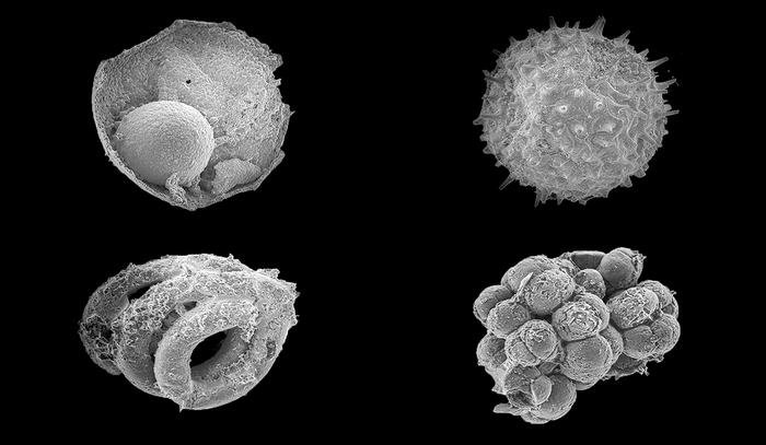
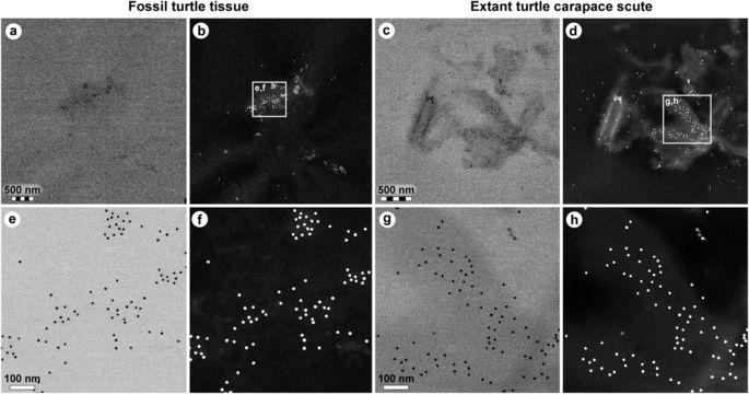
Mary Schweitzer, a biology graduate student at Montana State University’s Museum of the Rockies, was examining a thin section of Tyrranosaurus rex bone…when she noticed a series of peculiar structures. Round and tiny and nucleated, they were threaded through the bone like red blood cells in blood vessels. But blood cells in a dinosaur bone should have disappeared eons ago. “I got goose bumps,” recalls Schweitzer. “It was exactly like looking at a slice of modern bone. But, of course, I couldn’t believe it. I said to the lab technician: ‘The bones, after all, are 65 million years old. How could blood cells survive that long?'”2

Why was Schweitzer, an eyewitness who microscopically observed the insides of a T. rex bone, afraid to believe her own eyes? Isn’t empirical science all about observation? Furthermore, Morell reported, “Schweitzer has already extracted a molecule that might be dinosaur DNA.”

However, connective tissue ruins and degrades over time, such that DNA should not survive at all, even if the creature only lived 50,000 years ago.3 The existence of 65 million-year-old DNA is biochemically unthinkable. In other words, the old-earth evolutionary tale is clearly at odds with the fresh dinosaur bone evidence. How embarrassing to the academic establishment! This may be why ongoing dinosaur soft tissue discoveries are generally not broadcast through popular media channels.

Research Censorship

Evolutionary “damage control” is observed in the form of “chilling” (i.e., coerced) censorship of research, with severe consequences to those who “buck the system.” Consider the research flow chart pictured below describing the process of extracting dinosaur DNA. Note steps 7 and especially 8. Why must the research results be dismissed if the DNA extract doesn’t look like birds or crocodiles? The answer is evolutionary gatekeeping:

To make sure she’s liberated the right molecule, Schweitzer compares the extracted DNA sequences with those of hundreds of living organisms. If the sequence turns out to be similar to that of a known fungal gene, for example, she knows the sample has been contaminated.

That’s how DNA hunters know they’ve gone wrong. But how do they know when they’re on the right track, given that there are no living dinosaurs to provide a handy sample of DNA for comparison? The answer is that they rely on paleontological theory, which (according to most researchers) holds that dinosaurs and crocodiles came from the same stock, and that the dinosaurs’ only living descendants are birds. Therefore researchers look for DNA that is similar, but not identical, to DNA from these groups of organisms.4

In other words, only DNA research that provides dinosaur DNA sequences similar to those of birds and crocodiles is allowed. As the flowchart indicates, all other results are deemed anomalies that should be rejected as though they were known contaminants, like fungal genes. This approach is not observation-directed empirical research; this is assumption-driven, theory-dictated censorship–“science” falsely so-called.5

Extracting Dinosur DNA

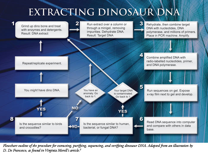
Extracting Dinosaur DNA

Coerced Spoliation of Evidence

This purposeful pattern of coerced concealment of the nonconforming DNA data from unfossilized dinosaur bones (labeled “an anomaly” on the chart) involves what courtroom lawyers and judges call “chilling” coercion and “spoliation of evidence”–inducing the concealment (and eventual destruction) of embarrassing information in order to prevent one’s opponent from using it at trial.

Whenever any kind of evidence is concealed, one immediately questions the spoliators’ motives for doing so. The intuitive answer is that they dislike what the information would reveal. Therefore, to spoliate evidence suggests that the spoliators’ argument or theory would be weakened, or embarrassed, by that evidence. This suggestion is so strong, forensically speaking, that it is treated as a rule of presumptive inference in law courts. In other words, if someone hides evidence in this way, the law presumes that the hidden evidence was damaging to the argument of the spoliator. The spoliator then bears the burden of proof to show otherwise.6

A kindred rule to the foregoing…is that the intentional spoliation or destruction of evidence relevant to a case raises a presumption that the evidence would have been unfavorable to the cause of the spoliator.…The deliberate destruction of evidence gives rise to the presumption that the matter destroyed is not favorable to the spoliator.7

This shows that the civil law courts understand the importance of evidence spoliation–it points to a willingness to conceal or otherwise suppress truth in order to advance a specific cause. The name Arthur Andersen comes to mind, as this accounting firm’s shredding of Enron documents hindered SEC investigators.8

Follow the Procedure, or Else

In suppressed dinosaur DNA research–which is a subset of the irrefutable, but hushed, dinosaur soft tissue discoveries–the same issue of evidence spoliation is relevant. Why? Because today’s dinosaur DNA controversy in particular, and today’s dinosaur “connective tissue” controversy in general, directly puts at issue the real age of the dinosaurs: Did they live millions of years ago, or in much more recent history on an earth inhabited by humans–descendants of Adam and Eve?9

How will anyone really know what dinosaur DNA sequences look like until uncensored data from dinosaur bones are published for public scrutiny? And how will such data be published at all if “embarrassing” research results are routinely discarded as anomalous, simply because they didn’t “look like chicken”? One way to acquire more reliable data in this case would be to repeat the DNA research across multiple labs, until consistent results emerge.

In fact, a similar approach was taken in 1994. The winners of the race to sequence dinosaur DNA were Scott Woodward and his colleagues, who published their results in Science.10 They extracted DNA from a purportedly well-preserved dinosaur bone. However, they were not rewarded for their victory. The sequence they discovered was not like birds or reptiles, but seemed unique.

These researchers decided not to follow the procedure outlined in the 1993 flowchart, which would have “told” them that what they found was an unacceptable “anomaly.” Since this 1994 DNA did not fit the evolutionary interpretive filter, the authors were raked over the academic coals. Moreover, the objections to their results were not based on conflicting research results, but appeared in editorials and reviews. As a result of the uproar from the scientific community, their dinosaur DNA sequence never became a permanent entry in any public database. In fact, since this very public academic flogging, no scientist has attempted to publish any dinosaur DNA research (resulting in “chilled” academic speech).

Interestingly, Schweitzer has never published any of her purported DNA research on dinosaur tissue, although she has published on tissue analyses and, recently, data on protein sequence. While the tissue analyses reported over the past decade are nearly impossible to dispute, this recently published dinosaur protein sequence from a T. rex came under extreme criticism and the data were highly questioned by peers as having been manipulated to produce close similarities with chicken and ostrich protein.11 Was this done as per the “paleontological theory and protocol” described in 1993?

Conclusion

The gatekeeping approach to ancient DNA research established as a protocol in 1993 is a product of dogmatic evolutionary theory. The 1994 results put the dogma to the test, with the result that:

  1. Ancient DNA, known to be unstable, was extracted from “80 million-year-old” bone.
  2. The sequence, though it showed evidence of decay, was no more bird-like than it was mammal-like.

The coerced suppression of the results by the evolutionary scientific community has dissuaded anyone else from publishing dinosaur DNA research that is not in line with evolutionary dictates. Such self-censorship “chills” empirical research, which prevents the public reporting of observable DNA sequences in order to insulate the larger story of particles-to-people evolution from cross-examination.

Where are the real scientists in dinosaur DNA research who refuse to kowtow to evolution’s gatekeepers?

References

  1. Thomas, B. 2009. Dinosaur Soft Tissue Issue Is Here to Stay. Acts & Facts. 38 (9): 18.
  2. Morell, V. 1993. Dino DNA: The Hunt and the Hype. Science. 261 (5118): 160.
  3. Ibid, 161. (This illustrates the thermodynamic maxim “as time increases, chemistry wins over biology.”)
  4. Difficulties With Dinosaur DNA, ibid, 161.
  5. 1 Timothy 6:20.
  6. See Wal-Mart Stores, Inc. v. Johnson, 106 S.W.3d 718, 46 Tex. Supr. Ct. J. 685 (Tex. 2003) (summarizing Texas jurisprudence regarding spoliation of evidence), citing Armory v. Delamirie, 1 Strange 505, 93 Engl. Rep. 664 (K.B. 1722) (illustrating how spoliation of evidence, as a legal problem, predates the USA’s existence).
  7. Quoting H.E. Butt Grocery Co. v. Bruner, 530 S.W.2d 340, 344 (Tex. Civ. App. – Waco, 1975, writ dismissed by agr’t), citing McCormick & Ray, TEXAS EVIDENCE (2nd ed.), Volume I, § 103, pages 141-142.
  8. See, e.g., In re Enron Corporation Securities, Derivative & “ERISA” Litigation, 2003 WL 25508889 (S.D. Tex. 2003) (discussing how Arthur Andersen accountants committed spoliation of evidence by shredding Enron documents to hinder the SEC’s investigation of Enron, etc.).
  9. There are indications that dinosaurs have lived within the last few thousands, and maybe even hundreds, of years. See Cooper, B. 1995. After the Flood. Chichester, UK: New Wine Press, 130-161, which documents and discusses historical records of human encounters with strange creatures during various centuries after Christ, involving detailed descriptions of wild animals that today would be called dinosaurs.
  10. Woodward, S. R., N. J. Weyand and M. Bunnell. 1994. DNA Sequence from Cretaceous Period Bone Fragments. Science. 266 (5188): 1229-1232.
  11. For more details, see Tomkins, J. 2009. Dinosaur Protein Sequences and the Dino-to-Bird Model. Acts & Facts. 38 (10): 12-14.

* Dr. Johnson is Special Counsel at ICR. Dr. Tomkins, ICR Research Associate, worked in academic research in genetics and genomics for 18+ years, 12 involving research in cloning and sequencing DNA from a wide variety of plants, animals and microbes. Mr. Thomas is Science Writer at the Institute for Creation Research.

Cite this article: Johnson, J. J. S., J. Tomkins and B. Thomas. 2009. Dinosaur DNA Research: Is the tale wagging the evidence? Acts & Facts. 38 (10): 4-6.

Source: http://www.icr.org

@ WFS,World Fossil Society,Riffin T Sajeev,Russel T Sajeev

WFS News:Dinosaur Gets Strange Diagnosis 78 Million Years After Its Death

@ WFS,World Fossil Society,Riffin T Sajeev,Russel T Sajeev

There weren’t any doctors when dinosaurs roamed the Earth, but one duck-billed dino has managed to get a diagnosis for its unusual joint condition about 78 million years after its death, thanks to a group of researchers who analyzed its strangely fused and pitted fossilized bones.

The adult duck-billed dinosaur, known as a hadrosaurid, had an inflammatory type of chronic arthritis, known as spondyloarthropathy, that attacks the spine and can cause the vertebrae to fuse together.

“This is the first occurrence of spondyloarthropathy in a hadrosaurid that we know [of],” said Darren Tanke, a senior fossil preparation technician at the Royal Tyrrell Museum of Palaeontology in Alberta, Canada, who is studying the fossils. [Photos: Duck-Billed Dinos Found in Alaska]

The duck-billed dinosaur's vertebrae were fused together because of its joint condition. Credit: Courtesy Royal Tyrrell Museum

 The duck-billed dinosaur’s vertebrae were fused together because of its joint condition.
                                                                  Credit: Courtesy Royal Tyrrell Museum

Researchers uncovered the unique fossils in 1988 from a bone bed — basically, an accumulation of bones from different dinosaurs in one location — at Milk River Ridge Reservoir, an artificial lake in southern Alberta.

“We were just doing a general collection of the bones along the shoreline there,” Tanke told Live Science. “The lake was lapping against the shoreline, exposing all these bones. They had to be collected right away, or they would have been destroyed.”

The scientists put the fossils into a plaster jacket, and worked on them over the years at the Royal Tyrell Museum, extracting horned and duck-billed dinosaur remains from the rock. Recently, a probe into the sample revealed a mysterious bone; it looked like the dinosaur’s sacrum — a bone in the lower back made of fused vertebrae that sits between the hip bones and the pelvis.

But this guess was wildly wrong, the researchers soon found.

“As [the fossil] became more and more cleaned up, we realized that the fusion of the vertebrae wasn’t the sacrum,” Tanke said. “It was fused because it was a pathology of the specimen, and the bones were not sacral vertebrae; they were dorsal vertebrae [in the middle of the back].”

It’s not uncommon to find fused duck-billed-dinosaur vertebrae, but these vertebrae are typically in the dinosaur’s tail, not in the middle of its spine, Tanke said.

Tanke excavated the rest of the duck-billed-dino specimen with extreme care, using an instrument that blasted air and an abrasive (in this case, baking soda) at the fossils to free them from the rock and clay.

The end result was unexpected: They found fused, pitted and textured vertebrae — a sign of spondyloarthropathy, said study co-researcher Dr. Bruce Rothschild, a professor in the School of Medicine at West Virginia University.

The condition likely impaired the dinosaur’s movement — a disadvantage if it was fleeing a predator, such as a tyrannosaur, Rothschild said. The ailment may have also made it difficult for the duck-billed creature to move around in everyday life and defend itself, he said.

Notice the pitted bone caused by the spondyloarthropathy. This bone would be smooth if the dinosaur didn't have this condition. Credit: Courtesy Royal Tyrrell Museum

Notice the pitted bone caused by the spondyloarthropathy. This bone would be smooth if the dinosaur didn’t have this condition.Credit: Courtesy Royal Tyrrell Museum

Evidence of spondyloarthropathy has also been found in other dinosaurs, including two species of horned dinosaur, six types of sauropod (a long-necked and long-tailed herbivorous giants) and two species of theropod (bipedal, mostly meat-eating dinosaurs, such as tyrannosaurs), the researchers said. The condition also affects mammals, including humans, Rothschild said. [The Strangest Medical Conditions]

There is no cure for the condition, but its symptoms in humans are often treated with the anti-inflammatory drug sulfasalazine (brand names Azulfidine and Sulfazine), Rothschild noted.

The research, which has yet to be published in a peer-reviewed journal, was presented Aug. 23 at the 2017 Society of Vertebrate Paleontology meeting in Calgary, Alberta.

Original article on Live Science.By Laura Geggel, Senior Writer.

@ WFS,World Fossil Society,Riffin T Sajeev,Russel T Sajeev

WFS News:Sea-level change and super storms; geologic evidence from the last interglacial (MIS 5e)

@ WFS,World Fossil Society,Riffin T Sajeev,Russel T Sajeev

While strong seasonal hurricanes have devastated many of the Caribbean and Bahamian islands this year, geologic studies on several of these islands illustrate that more extreme conditions existed in the past. A new analysis published in Marine Geology shows that the limestone islands of the Bahamas and Bermuda experienced climate changes that were even more extreme than historical events. In the interest of our future world, scientists must seek to understand the complexities of linked natural events and field observations that are revealed in the geologic record of past warmer climates.

In Bermuda and the Bahamas, the geology of the last interglacial (LIG; approximately 120,000 years ago) is exquisitely preserved in nearly pure carbonate sedimentary rocks. A record of superstorms and changing sea levels is exposed in subtidal, beach, storm, and dune deposits on multiple islands. Extensive studies by the authors over the past decades on these islands have documented stratigraphic, sedimentologic, and geomorphic evidence of major oceanic and climatic disruptions at the close of the last interglacial.

The image on the left shows eolian (lower) and runup bedding (upper) exposed in a roadcut on Old Land Road on Great Exuma Island (road elevation +23 meters). On the right are thick beds with fenestral porosity, or 'beach bubbles,' showing that massive waves ran up over older dunes exposed in a roadcut on Suzy Turn Road along the Atlantic Ocean east side of Providenciales, Turks and Caicos Islands, BWI. Credit: Marine Geology

The image on the left shows eolian (lower) and runup bedding (upper) exposed in a roadcut on Old Land Road on Great Exuma Island (road elevation +23 meters). On the right are thick beds with fenestral porosity, or ‘beach bubbles,’ showing that massive waves ran up over older dunes exposed in a roadcut on Suzy Turn Road along the Atlantic Ocean east side of Providenciales, Turks and Caicos Islands, BWI. Credit: Marine Geology

Dr. Paul J. Hearty, a retired Associate Professor at the University of North Carolina at Wilmington, and Dr. Blair. R. Tormey, a Coastal Research Scientist at Western Carolina University conducted an invited review of published findings. It demonstrates that during a global climate transition in the late last interglacial, also known as marine isotope substage 5e (MIS 5e), abrupt multi-meter sea-level changes occurred. Concurrently, coastlines of the Bahamas and Bermuda were impacted by massive storms generated in the North Atlantic Ocean, resulting in a unique trilogy of wave-transported deposits: megaboulders, chevron-shaped, storm-beach ridges, and runup deposits on high dune ridges.

While perhaps more mundane than the megaboulders (found only locally on Eleuthera), the sedimentological structures found within chevron ridge and runup deposits across islands throughout the Bahamas and Bermuda point to frequent and repeated inundation by powerful storm waves, in some locations leaving storm deposits tens of meters above sea level.

During the last interglacial, sea levels were about 3-9 meters higher than they are now. The geologic evidence indicates that the higher sea-levels were accompanied by intense “superstorms,” which deposited giant wave-transported boulders at the top of cliffed coastlines, formed chevron-shaped, storm beach ridges in lowland areas, and left wave runup deposits on older dunes more than 30 meters above sea level. These events occurred at a time of only slightly warmer global climate and CO2 (about 275 ppm) was much lower than today.

The authors emphasize “the LIG record reveals that strong climate forcing is not required to yield major impacts on the ocean and ice caps.” In our industrial world, rapidly increasing atmospheric CO2 has surpassed 400 ppm, levels not achieved since the Pliocene era about 3 million years ago, while global temperature has increased nearly 1 °C since the 1870s. Today, ice sheets are melting, sea level is rising, oceans are warming, and weather events are becoming more extreme.

Drs. Hearty and Tormey conclude that with the greatly increased anthropogenic CO2 forcing at rates unmatched in nature, except perhaps during global extinction events, dramatic change is certain. They caution that, “Our global society is producing a climate system that is racing forward out of humanity’s control into an uncertain future. If we seek to understand the non-anthropogenic events of the last interglaciation, some of the consequences of our unchecked forward speed may come more clearly into focus…a message from the past; a glimpse into the future.”

@ WFS,World Fossil Society,Riffin T Sajeev,Russel T Sajeev

Ref: P.J. Hearty, B.R. Tormey. Sea-level change and superstorms; geologic evidence from the last interglacial (MIS 5e) in the Bahamas and Bermuda offers ominous prospects for a warming Earth. Marine Geology, 2017; 390: 347 DOI: 10.1016/j.margeo.2017.05.009

WFS News:Fossil discovery in Tanzania reveals ancient bobcat-sized carnivore

@ WFS,World Fossil Society,Riffin T Sajeev,Russel T Sajeev

Paleontologists working in Tanzania have identified a new species of hyaenodont, a type of extinct meat-eating mammal. The study is published today, National Fossil Day, in the journal PLOS ONE and funded by the National Science Foundation (NSF).

After the extinction of the non-avian dinosaurs 66 million years ago, hyaenodonts were the main predators on the African continent. The newly discovered animal is called Pakakali rukwaensis, the name derived from the Swahili term “pakakali,” meaning “fierce cat,” and “rukwaensis,” the word for the Rukwa Rift region of the Great Rift Valley in southwestern Tanzania.

A closer look at the bobcat-like fossil animal uncovered in Tanzania. Credit: Matthew Borths

A closer look at the bobcat-like fossil animal uncovered in Tanzania.
Credit: Matthew Borths

Between 23 and 25 million years ago, newcomers arrived in Africa — the first relatives of modern dogs, cats and hyenas — where they coexisted with hyaenodonts for millions of years. But eventually, hyaenodonts went extinct.

“The shift from hyaenodonts to modern carnivores in Africa is like a controlled experiment,” says study co-author Matthew Borths of Ohio University.

“We start with only hyaenodonts. Then the relatives of cats and dogs arrive. They coexist for a few million years, then the hyaenodonts are driven to extinction and we’re left with ‘The Lion King.’ With Pakakali, we can start to unravel that extinction. Were the lineages competing? Were they adapting differently to a drier, more open landscape?”

The new fossil helps researchers unravel extinction dynamics for predatory mammals stalking African ecosystems of that long-ago time.

“This new carnivore, discovered in Tanzania sediment deposits dating from 25 million years ago, provides new information about the transition of carnivores in older ecosystem types to carnivores in today’s African ecosystems,” says Judy Skog, program director in NSF’s Division of Earth Sciences, which funded the research.

Paleontologists at work in Tanzania on research that led to the find of the new carnivore species. Credit: Nancy Stevens

Paleontologists at work in Tanzania on research that led to the find of the new carnivore species.
Credit: Nancy Stevens

The new hyaenodont species was discovered in the same 25 million-year-old rocks as the oldest fossil evidence of the split between Old World monkeys and apes. At that time, the ecosystem was undergoing dramatic climate and tectonic upheavals as Africa collided with Eurasia and the modern East African Rift System formed.

The fossil gives paleontologists a glimpse of hyaenodont anatomy before modern carnivores invaded the continent, revealing that Pakakali was about the size of a bobcat.

Based on the findings of the study, hyaenodonts may have been pushed to become more specialized meat-eaters due to competition from other species. That dietary specialization may have made hyaenodonts more vulnerable to extinction in the changing African ecosystem by leaving them with fewer food choices.

Pakakali was discovered by an international team of scientists from the United States, Australia and Tanzania as part of the Rukwa Rift Basin Project (RRBP), an interdisciplinary collaboration examining the development of the modern African ecosystem. In more than a decade of exploration, RRBP researchers have described the habitat Pakakali called home along with many other animals that occupied the ecosystem.

“The environment containing Pakakali reveals a fascinating window into extinction,” says Nancy Stevens, co-author of the study and a paleontologist at Ohio University. “It highlights the vulnerability of carnivorous species to rapid environmental change, a topic we are grappling with on the African continent today.”

Source: The National Science Foundation (NSF):News Release 17-101

@ WFS,World Fossil Society,Riffin T Sajeev,Russel T Sajeev