WFS News: Foxaspis novemura: A new fossilized galeaspid species

@WFS,World Fossil Society, Athira,Riffin T Sajeev,Russel T Sajeev

A new fossilized galeaspid speciesFoxaspis novemura, has been discovered in China, featuring a unique nine-rayed caudal fin. The species was found to be an active swimmer with a faster cruising speed than its relatives.

Researchers have discovered a new fossilized galeaspid species with “nine tails” in south China’s Guangxi Zhuang Autonomous Region. This is the world’s first galeaspid fossil with a well-preserved tail.

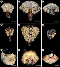
Photo shows a complete headshield articulated with body and tail preserved together with a complete arthrodiran fish. Credit: Chinese Academy of Sciences

Photo shows a complete headshield articulated with body and tail preserved together with a complete arthrodiran fish. Credit: Chinese Academy of Sciences

According to the researchers from the Institute of Vertebrate Paleontology and Paleoanthropology under the Chinese Academy of Sciences, they named the new species as Foxaspis novemura because its caudal fin is comprised of nine ray-like scale-covered digitations, just like that of the Nine-Tailed Fox, a mythical animal from an ancient literature “Shan Hai Jing” (Classic of Mountains and Seas).

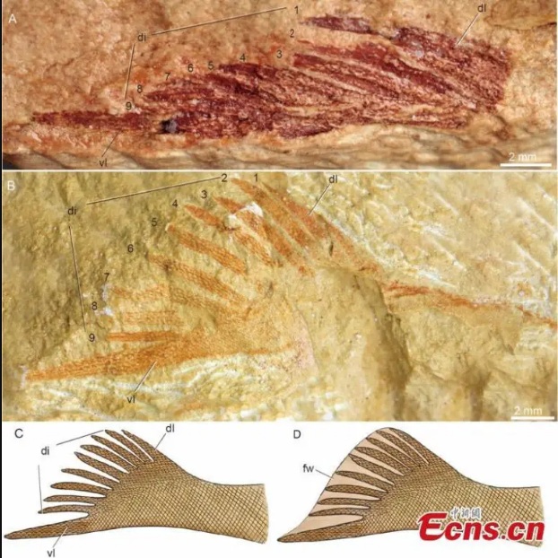
Photo shows an exceptionally preserved fossil of “nine tails.” Credit: Chinese Academy of Sciences

Photo shows an exceptionally preserved fossil of “nine tails.” Credit: Chinese Academy of Sciences

The fossil specimen completely preserves the caudal fin in both folded and flared states, revealing its morphological details to the greatest extent, according to Gai Zhikun, a research professor from the institute.

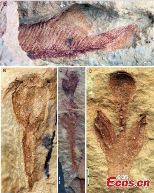
Photo shows the exceptionally preserved fossils of fishes and plants associated with Foxaspis. Credit: Chinese Academy of Sciences

Photo shows the exceptionally preserved fossils of fishes and plants associated with Foxaspis. Credit: Chinese Academy of Sciences

“We discovered that galeaspids may be active swimmers, and can make good use of muscle contraction to control the contact area between tail and water flow, thus generating different thrust forces,” he said.

The team then analyzed the swimming speed of the geometrical morphology of the tail and showed that galeaspid’s cruising speed was even faster than that of their more derived jawless and jawed relatives.

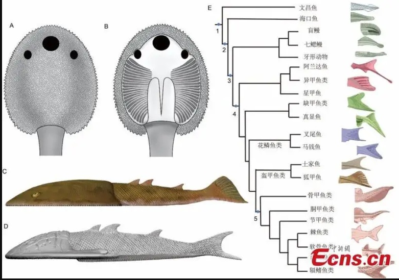
Life restoration and caudal fin evolution of the “nine tails.” Credit: Chinese Academy of Sciences

Life restoration and caudal fin evolution of the “nine tails.” Credit: Chinese Academy of Sciences

The researchers published their study results in the journal National Science Review.

Reference: “Postcranial disparity of galeaspids and the evolution of swimming speeds in stem-gnathostomes” by Zhikun Gai, Xianghong Lin, Xianren Shan, Humberto G Ferrón and Philip C J Donoghue, 27 February 2023, National Science Review.DOI: 10.1093/nsr/nwad050

@WFS,World Fossil Society, Athira,Riffin T Sajeev,Russel T Sajeev

WFS News: Fossil evidence of tylosis formation in Late Devonian plants

@WFS,World Fossil Society, Athira,Riffin T Sajeev,Russel T Sajeev

An international team of scientists, co-led by Dr Carla J. Harper, Assistant Professor in Botany in the School of Natural Sciences at Trinity, has discovered the oldest evidence of plant self-defence in wood in a 360-million-year-old fossil from south-eastern Ireland.

Plants can protect their wood from infection and water loss by forming special structures called “tyloses.” These prevent bacterial and fungal pathogens from getting into the heartwood of living trees and damaging it. However, it was not previously known how early in the evolution of plants woody species became capable of forming such defences.

Published today in Nature Plants is the oldest evidence of tylosis formation from Late Devonian (360-million-year-old) fossil wood from the Hook Head Peninsula area, Co. Wexford, Ireland.

These plants lived well before the time of the dinosaurs or even flying insects. They formed the first primeval forests, when plants ruled the continents, accompanied by microorganisms, fungi, and early relatives of spiders, millipedes and centipedes.

Dr Harper and her team, including Dr Anne-Laure Decombeix (CNRS, France), Dr Cyrille Prestianni (Uni. Liège, Belgium), Trinity Botany PhD student Thibault Durieux (co-advised by Harper and Decombeix), Merlin Ramel (INRAe, France), and Prof Michael Krings (BSGP, Munich, Germany and Trinity SNS Visiting Research Associate), discovered tyloses in the fossilised wood of an extinct group of plants known as the Archaeopteridalean progymnosperms. These plants are particularly important as they were the first trees to resemble those we see today, with a large woody trunk, branches, and complex root systems.

The team has now discovered that these primitive trees were also able to form tyloses to protect their wood. What is particularly exciting is that Ireland is one of the few places in the world where such details can be observed in plants from this remote time period. This means that the fossils from Co. Wexford give unique insights into this important period in plant evolution.

Dr Harper said: “Fossil wood is an example of an anatomically preserved fossil: plant remains that have been infiltrated by a water rich in minerals, preserving their tissues in three dimensions. These fossils allow us to study very fine details of extinct plant anatomy, down to the cellular level. This type of preservation, in general, is rare but occurs in certain fossil deposits in Ireland.

“Continuing fieldwork in Irish Devonian localities will yield new fossils that will increase our understanding of the diversity and biology of extinct plants. Overall, Ireland’s rich plant fossil history — an untapped resource — plays a key role in answering exciting research questions and raises many more.”

Ireland has long been known as the Emerald Isle due its famous rolling green hills — but such discoveries help us to understand how and when this “greening” began.

Dr Harper said: “By studying these fossil plants and their past environments, we can get powerful insights into the history of plant physiological processes that still occur today, and into both current and future ecosystems of Ireland and the world.”

  1. Anne-Laure Decombeix, Carla J. Harper, Cyrille Prestianni, Thibault Durieux, Merlin Ramel, Michael Krings. Fossil evidence of tylosis formation in Late Devonian plantsNature Plants, 2023; DOI: 10.1038/s41477-023-01394-0
Trinity College Dublin. “360-million-year-old Irish fossil provides oldest evidence of plant self-defense in wood.” ScienceDaily. ScienceDaily, 21 April 2023. <www.sciencedaily.com/releases/2023/04/230421134352.htm>.
@WFS,World Fossil Society, Athira,Riffin T Sajeev,Russel T Sajeev

WFS News: World’s oldest preserved brain found in prehistoric fish fossil

@WFS,World Fossil Society, Athira,Riffin T Sajeev,Russel T Sajeev

The skull fossil is the only known specimen of its species, so only non-destructive techniques were used during the study

The skull fossil is the only known specimen of its species, so only non-destructive techniques were used during the study

The “oldest example of a well-preserved vertebrate brain” has been identified in a 319 million-year-old fish fossil.

The fossilised Coccocephalus wildi was found in a coal mine in Lancashire more than a century ago and had been sitting in the archives of Manchester Museum.

Research by teams from universities in Birmingham and Michigan found its skull contained a brain and cranial nerves.

Palaeontologist Dr Sam Giles said the “unexpected find” gave a “startling insight” into brain evolution.

The fossil of the now-extinct fish was originally recovered from a layer of soapstone in the roof of the Lancashire mine and was first scientifically described in 1925.

Though only its skull was recovered, scientists from the University of Birmingham (UoB) and the University of Michigan (UoM) believe it would have been 6 to 8ins (15 to 20cm) long and was probably a carnivore.

A UoB representative said soft tissues like brains normally decay quickly and “very rarely fossilise”, but when this fish died, it was “probably quickly buried in sediments with little oxygen present” as such environments “can slow the decomposition of soft body parts”.

They said the skull fossil was the only known specimen of its species, so only non-destructive techniques were used during the study.

They added that the research team had not been looking for a brain when examining the skull fossil, but found the distinct object which had features found in vertebrate brains, such as bilateral symmetry and hollow spaces.

Scans of the fossil revealed a brain and cranial nerves, researchers said

Scans of the fossil revealed a brain and cranial nerves, researchers said

Dr Giles said the “unexpected find of a three-dimensionally preserved vertebrate brain gives us a startling insight into the neural anatomy of ray-finned fish”.

“It tells us a more complicated pattern of brain evolution than suggested by living species alone, allowing us to better define how and when present day bony fishes evolved,” she added.

UoM researcher Rodrigo Figueroa said the “superficially unimpressive and small fossil [not only] shows us the oldest example of a fossilised vertebrate brain, but it also shows that much of what we thought about brain evolution from living species alone will need reworking”.

Source:  Article in BBC NEWS.com dtd 2/3/2023

@WFS,World Fossil Society, Athira,Riffin T Sajeev,Russel T Sajeev

WFS News: Australia’s most intact Cooyoo australis fossil discovered in Richmond with specimen in its belly

@WFS,World Fossil Society, Athira,Riffin T Sajeev,Russel T Sajeev

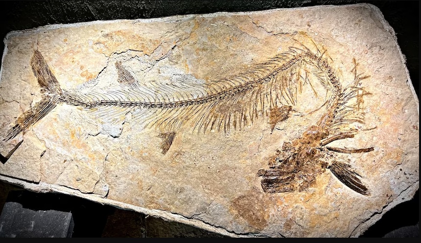
An intact cooyoo fish fossil, about 1.6 metres long, has been discovered in Richmond.(Supplied: Kronosaurus Korner)

An intact cooyoo fish fossil, about 1.6 metres long, has been discovered in Richmond.(Supplied: Kronosaurus Korner)

The fossil of a predator fish that existed more than 100 million years ago has been discovered in north-west Queensland.

Experts believe it is one Australia’s most intact fossils of the Cooyoo australis fish — a large carnivore that once swam Queensland’s inland Eromanga Sea.

The rare find was stumbled upon by a Northern Territory couple who had been digging at one of the public fossicking sites near the Kronosaurus Korner museum in Richmond — an area renowned for its significant fossil discoveries.

But the quality of the 1.6-metre specimen is not the only thing that has archaeologists excited.

“There are two extremely special things about this fish,” Kronosaurus Korner founder and chairman Rob Ievers said.

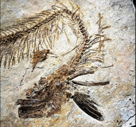
“This thing has all its ribs, all its vertebrae … a complete head with large jaws, and a mouthful of sharp teeth.”

The cooyoo was of such high quality, curators were able to see the contents of its stomach.

“This is most interesting because you don’t find this very often at all, we can see what this fish had for its last meal,” Mr Ievers said.

It turns out fish was on the menu for this cooyoo.

The head of the cooyoo fossil would have been lined with sharp teeth.(Supplied: Kronosaurus Korner)

The head of the cooyoo fossil would have been lined with sharp teeth.(Supplied: Kronosaurus Korner)

“When you get a close-up view of the specimen, you can see the little vertebrae, you can see a small fish head and some other remains of a fish in the area where the cooyoo’s stomach would have been,” Mr Ievers said.

He said the tidal nature of the Eromanga Sea and the presence of other carnivores meant it was rare to find a whole specimen.

“Because, when these things died, this sea was tidal so there was movement and these fish got displaced through the natural process of rotting and with other animals feeding off them,” Mr Ievers said.

A beastly sight

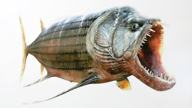
Cooyoo had large conical teeth within deep jaws, used for swallowing prey whole.(Supplied: Kronosaurus Korner)

Cooyoo had large conical teeth within deep jaws, used for swallowing prey whole.(Supplied: Kronosaurus Korner)

With a gaping underbite lined in conical teeth and a bull-dog head, the Cooyoo australis had a face only a mother could love.

Several sets of fins, a long body, and forked tail would have helped it torpedo through the water, threatened only by large marine reptiles and sharks.

One of the largest semi-articulated cooyoo specimens was discovered by Gary and Barb Flewelling and Anthony Saffioti in 2011 in Richmond.

The 2.5-metre long fossil nicknamed “Wandah” is currently on display at Kronosaurus Korner.

Source: Article by By ABC North West Qld /Zara Margolis and Larissa Waterson

@WFS,World Fossil Society, Athira,Riffin T Sajeev,Russel T Sajeev


WFS News: Fossil site reveals giant arthropods dominated the seas 470 million years ago

@WFS,World Fossil Society, Athira,Riffin T Sajeev,Russel T Sajeev

Discoveries at a major new fossil site in Morocco suggest giant arthropods — relatives of modern creatures including shrimps, insects and spiders — dominated the seas 470 million years ago.

Early evidence from the site at Taichoute, once undersea but now a desert, records numerous large “free-swimming” arthropods.

More research is needed to analyse these fragments, but based on previously described specimens, the giant arthropods could be up to 2m long.

An international research team say the site and its fossil record are very different from other previously described and studied Fezouata Shale sites from 80km away.

(a) Stratigraphic column for the Lower Ordovician Fezouata Shale and Zini formations including the Taichoute locality divided into three fossil assemblages. The red arrow in (a) indicates an accumulation level of brachiopods and bryozoans at the top of the Taichoute section. (b) Zoom on the Zagora Region where the Fezouata Shale was discovered. (c) Taichoute is the most distal site in the depositional environment of the Fezouata Shale. (d) Concretion from the Zagora region. (e) A large lobe from the Fezouata Shale in the upper part of the succession (a3) and interpreted as a density-flow deposit.

(a) Stratigraphic column for the Lower Ordovician Fezouata Shale and Zini formations including the Taichoute locality divided into three fossil assemblages. The red arrow in (a) indicates an accumulation level of brachiopods and bryozoans at the top of the Taichoute section. (b) Zoom on the Zagora Region where the Fezouata Shale was discovered. (c) Taichoute is the most distal site in the depositional environment of the Fezouata Shale. (d) Concretion from the Zagora region. (e) A large lobe from the Fezouata Shale in the upper part of the succession (a3) and interpreted as a density-flow deposit.

They say Taichoute (considered part of the wider “Fezouata Biota”) opens new avenues for paleontological and ecological research.

“Everything is new about this locality — its sedimentology, paleontology, and even the preservation of fossils — further highlighting the importance of the Fezouata Biota in completing our understanding of past life on Earth,” said lead author Dr Farid Saleh, from the University of Lausanne and and Yunnan University.

Dr Xiaoya Ma, from the University of Exeter and Yunnan University, added: “While the giant arthropods we discovered have not yet been fully identified, some may belong to previously described species of the Fezouata Biota, and some will certainly be new species.

“Nevertheless, their large size and free-swimming lifestyle suggest they played a unique role in these ecosystems.”

The Fezouata Shale was recently selected as one of the 100 most important geological sites worldwide because of its importance for understanding the evolution during the Early Ordovician period, about 470 million years ago.

Fossils discovered in these rocks include mineralised elements (eg shells), but some also show exceptional preservation of soft parts such as internal organs, allowing scientists to investigate the anatomy of early animal life on Earth.

Animals of the Fezouata Shale, in Morocco’s Zagora region, lived in a shallow sea that experienced repeated storm and wave activities, which buried the animal communities and preserved them in place as exceptional fossils.

However, nektonic (or free-swimming) animals remain a relatively minor component overall in the Fezouata Biota.

The new study reports the discovery of the Taichoute fossils, preserved in sediments that are a few million years younger than those from the Zagora area and are dominated by fragments of giant arthropods.

Fossils from the Taichoute locality (a1: A–C, a2: D–F, a3: G–J). (a) The gastropod Lesueurilla prima. (b) The solutan echinoderm Plasiacystis mobilis. (c) The graptolite Baltograptus gr. deflexus. (d–e) Giant euarthropod carapaces indicated with white arrows. (d) Carapace likely belonging to previously documented bivalved arthropods from the Fezouata Biota59. (e) Two incomplete but tapering carapaces (left and center) adjacent to a structure that bears possible resemblance to a block of radiodont setal blades (right), consisting of a series of parallel elongated blades a few millimetres wide separated by slight changes in sediment level and/or by intervening sediment, with an overall tapering outline, similar to the setal blade blocks of hurdiids such as Hurdia from the Burgess Shale60. (f) The multiramous graptolite Holograptus sp. (g) The calymenid trilobite Colpocoryphe cf. thorali. (h) The illaenid trilobite Ectillaenus? sp. (i) The calymenid trilobite Neseuretus cf. attenuatus (blue arrow) and Lichidae gen. indet. (to the left of the blue arrow). (j) A dalmanitid trilobite, subfamily Zeliszkellinae. (k) Accumulations of specimens of a new genus of orthidine brachiopod and bryozoans (white arrow) on top of a3. Scale bars = 5 mm in a, f, g, h, i, j; 10 mm in b, c and k; 25 mm in d; 50 mm in e. By order from a to j: AA.TAI2.OI.9; ML20-259,357; AA.TAI3.OI.2; AA.TAI6.OI.1; AA.TAI6.OI.2; AA.TAI6.OI.6; AA.TAI13.OI.1; AA.TAI13.OI.2; AA.TAI13.OI.3; AA.TAI13.OI.4; AA.TAI14.OI.1. All specimens are housed in the Marrakech Collections of the Cadi Ayyad University.

Fossils from the Taichoute locality (a1: A–C, a2: D–F, a3: G–J). (a) The gastropod Lesueurilla prima. (b) The solutan echinoderm Plasiacystis mobilis. (c) The graptolite Baltograptus gr. deflexus. (d–e) Giant euarthropod carapaces indicated with white arrows. (d) Carapace likely belonging to previously documented bivalved arthropods from the Fezouata Biota59. (e) Two incomplete but tapering carapaces (left and center) adjacent to a structure that bears possible resemblance to a block of radiodont setal blades (right), consisting of a series of parallel elongated blades a few millimetres wide separated by slight changes in sediment level and/or by intervening sediment, with an overall tapering outline, similar to the setal blade blocks of hurdiids such as Hurdia from the Burgess Shale60. (f) The multiramous graptolite Holograptus sp. (g) The calymenid trilobite Colpocoryphe cf. thorali. (h) The illaenid trilobite Ectillaenus? sp. (i) The calymenid trilobite Neseuretus cf. attenuatus (blue arrow) and Lichidae gen. indet. (to the left of the blue arrow). (j) A dalmanitid trilobite, subfamily Zeliszkellinae. (k) Accumulations of specimens of a new genus of orthidine brachiopod and bryozoans (white arrow) on top of a3. Scale bars = 5 mm in a, f, g, h, i, j; 10 mm in b, c and k; 25 mm in d; 50 mm in e. By order from a to j: AA.TAI2.OI.9; ML20-259,357; AA.TAI3.OI.2; AA.TAI6.OI.1; AA.TAI6.OI.2; AA.TAI6.OI.6; AA.TAI13.OI.1; AA.TAI13.OI.2; AA.TAI13.OI.3; AA.TAI13.OI.4; AA.TAI14.OI.1. All specimens are housed in the Marrakech Collections of the Cadi Ayyad University.

“Carcasses were transported to a relatively deep marine environment by underwater landslides, which contrasts with previous discoveries of carcass preservation in shallower settings, which were buried in place by storm deposits,” said Dr Romain Vaucher, from the University of Lausanne.

Professor Allison Daley, also from the University of Lausanne, added: “Animals such as brachiopods are found attached to some arthropod fragments, indicating that these large carapaces acted as nutrient stores for the seafloor dwelling community once they were dead and lying on the seafloor.”

Dr Lukáš Laibl, from the Czech Academy of Sciences, who had the opportunity to participate in the initial fieldwork, said: “Taichoute is not only important due to the dominance of large nektonic arthropods.

“Even when it comes to trilobites, new species so far unknown from the Fezouata Biota are found in Taichoute.”

Dr Bertrand Lefebvre, from the University of Lyon, who is the senior author on the paper, and who has been working on the Fezouata Biota for the past two decades, concluded: “The Fezouata Biota keeps surprising us with new unexpected discoveries.”

The paper, published in the journal Scientific Reports, is entitled: “New fossil assemblages from the Early Ordovician Fezouata Biota.”

Journal Reference:

  1. Farid Saleh, Romain Vaucher, Muriel Vidal, Khadija El Hariri, Lukáš Laibl, Allison C. Daley, Juan Carlos Gutiérrez-Marco, Yves Candela, David A. T. Harper, Javier Ortega-Hernández, Xiaoya Ma, Ariba Rida, Daniel Vizcaïno, Bertrand Lefebvre. New fossil assemblages from the Early Ordovician Fezouata BiotaScientific Reports, 2022; 12 (1) DOI: 10.1038/s41598-022-25000-z

Source: Science Daily

@WFS,World Fossil Society, Athira,Riffin T Sajeev,Russel T Sajeev

WFS News: Geobiologists shine new light on Earth’s first known mass extinction event 550 million years ago

@WFS,World Fossil Society, Athira, Riffin T Sajeev,Russel T Sajeev

A new study by Virginia Tech geobiologists traces the cause of the first known mass extinction of animals to decreased global oxygen availability, leading to the loss of a majority of animals present near the end of the Ediacaran Period some 550 million years ago.

The research spearheaded by Scott Evans, a postdoctoral researcher in the Department of Geosciences, part of the Virginia Tech College of Science, shows this earliest mass extinction of about 80 percent of animals across this interval. “This included the loss of many different types of animals, however those whose body plans and behaviors indicate that they relied on significant amounts of oxygen seem to have been hit particularly hard,” Evans said. “This suggests that the extinction event was environmentally controlled, as are all other mass extinctions in the geologic record.”

Evans’ work was published Nov. 7 in the Proceedings of the National Academy of Sciences, a peer-reviewed journal of the National Academy of Sciences. The study was co-authored by Shuhai Xiao, also a professor in the Department of Geosciences, and several researchers led by Mary Droser from the University of California Riverside’s Department of Earth and Planetary Sciences, where Evans earned his master’s degree and Ph.D.

“Environmental changes, such as global warming and deoxygenation events, can lead to massive extinction of animals and profound disruption and reorganization of the ecosystem,” said Xiao, who is an affiliated member of the Global Change Center, part of the Virginia Tech Fralin Life Sciences Institute. “This has been demonstrated repeatedly in the study of Earth history, including this work on the first extinction documented in the fossil record. This study thus informs us about the long-term impact of current environmental changes on the biosphere.”

What exactly caused the drop in global oxygen? That’s still up for debate. “The short answer to how this happened is we don’t really know,” Evans said. “It could be any number and combination of volcanic eruptions, tectonic plate motion, an asteroid impact, etc., but what we see is that the animals that go extinct seem to be responding to decreased global oxygen availability.”

The study by Evans and Xiao is timelier than one would think. In an unconnected study, Virginia Tech scientists recently found that anoxia, the loss of oxygen availability, is affecting the world’s fresh waters. The cause? The warming of waters brought on by climate change and excess pollutant runoff from land use. Warming waters diminish fresh water’s capacity to hold oxygen, while the breakdown of nutrients in runoff by freshwater microbes gobbles up oxygen.

“Our study shows that, as with all other mass extinctions in Earth’s past, this new, first mass extinction of animals was caused by major climate change — another in a long list of cautionary tales demonstrating the dangers of our current climate crisis for animal life,” said Evans, who is an Agouron Institute Geobiology fellow.

Some perspective: The Ediacaran Period spanned roughly 96 million years, bookended on either side by the end of Cryogenian Period — 635 million years ago — and the beginning of the Cambrian Period — 539 million years ago. The extinction event comes just before a significant break in the geologic record, from the Proterozoic Eon to the Phanerozoic Eon.

There are five known mass extinctions that stand out in the history of animals, the “Big Five,” according to Xiao, including the Ordovician-Silurian Extinction (440 million years ago), the late Devonian Extinction (370 million years ago), the Permian-Triassic Extinction (250 million years ago), the Triassic-Jurassic Extinction (200 million years ago), and the Cretaceous-Paleogene Extinction (65 million years ago).

“Mass extinctions are well recognized as significant steps in the evolutionary trajectory of life on this planet,” Evans and team wrote in the study. Whatever the instigating cause of the mass extinction, the result was multiple major shifts in environmental conditions. “Particularly, we find support for decreased global oxygen availability as the mechanism responsible for this extinction. This suggests that abiotic controls have had significant impacts on diversity patterns throughout the more than 570 million-year history of animals on this planet,” the authors wrote.

Fossil imprints in rock tell researchers how the creatures that perished in this extinction event would have looked. And they looked, in Evans’ words, “weird.”

“These organisms occur so early in the evolutionary history of animals that in many cases they appear to be experimenting with different ways to build large, sometimes mobile, multicellular bodies,” Evans said. “There are lots of ways to recreate how they look, but the take-home is that before this extinction the fossils we find don’t often fit nicely into the ways we classify animals today. Essentially, this extinction may have helped pave the way for the evolution of animals as we know them.”

The study, like scores of other recent publications, came out of the COVID-19 pandemic. Because Evans, Xiao, and their team couldn’t get access to the field, they decided to put together a global database based mostly on published records to test ideas about changing diversity. “Others had suggested that there might be an extinction at this time, but there was a lot of speculation. So we decided to put together everything we could to try and test those ideas.” Evans said. Much of the data used in the study was collected by Droser and several graduate students from the University of California Riverside.

ournal Reference:

  1. Scott D. Evans, Chenyi Tu, Adriana Rizzo, Rachel L. Surprenant, Phillip C. Boan, Heather McCandless, Nathan Marshall, Shuhai Xiao, Mary L. Droser. Environmental drivers of the first major animal extinction across the Ediacaran White Sea-Nama transitionProceedings of the National Academy of Sciences, 2022; 119 (46) DOI: 10.1073/pnas.2207475119
Source: www.sciencedaily.com/releases/2022/11/221107153524.htm
@WFS,World Fossil Society, Athira, Riffin T Sajeev,Russel T Sajeev

WFS News: 2-Billion-Year-Old Microbe Fossils found

@WFS,World Fossil Society,Athira,Riffin T Sajeev,Russel T Sajeev

Not all fossils are remnants from ferocious dinos. Some of them are teeny-tiny blobs.

Scientists recently discovered some of these blobs in the form of 2.5-billion-year-old fossils of primitive bacteria. These ancient microbes are likely cyanobacteria, but they are unusually large and have weird shapes protruding from them, said Andrew Czaja, an associate professor at the University of Cincinnati, who presented his findings on Wednesday (June 26) at the Astrobiology Science Conference.

If these fossils really are cyanobacteria, they could be some of the primitive organisms, or their ancestors, that helped transform our atmosphere by pumping it with oxygen. But not everyone is convinced.

The newly discovered fossils come from a period 100 million to 200 million years before the Great Oxidation Event — when our atmosphere went from having no oxygen to having a little bit.

“This is a very important time in Earth’s history, both in terms of the evolution of the Earth but also the evolution of life,” Czaja told Live Science.

Yet, “we don’t actually have many instances of fossils from this time period.” Czaja said. Czaja said he knew of only four cases in the literature of microfossils dating to between 2.5 billion and 2.7 billion years ago.

Czaja was exploring in South Africa when he happened upon a cool-looking rock, called a stromatolite, which is made up of layers of limestone and sediments left behind by cyanobacteria.

He brought it home to show during his classes, but it turned out the rock was chock-full of microfossils. Andrea Corpolongo, a doctoral student also at the University of Cincinnati, then began to analyze the rock under a microscope. The fossils turned out to be hollow spheres made of an organic compound called kerogen. Some of those spheres were oblong and some had weird protrusions coming off them.

The researchers don’t know exactly what kind of microbes they’re looking at, but because these fossils were found in the stromatolite, they may be ancient cyanobacteria. Yet some of them are bigger than any cyanobacteria we have today.

Nowadays, most cyanobacteria range from 5 to 10 microns, with the largest of these creatures measuring 60 microns, Czaja said. These ancient microbe fossils have a wide range of sizes, but most are above the average size of today’s cyanobacteria and some are up to 100 microns across.

They also don’t know why some of them have weird protrusions, which at first glance seem to be a type of “budding,” orf asexual reproduction in which a part of an organism splits off to become a new organism. Nowadays, cyanobacteria don’t bud and so “i’m not really claiming it’s budding, but it does look like that,” he said.

Emily Kraus, a doctoral student at the Colorado School of Mines, wasn’t convinced.

“What he says are microfossils are very large,” said Kraus, who wasn’t involved with the new research. “They’re larger than cells and cyanobacteria, which don’t look like that, so I wasn’t terribly convinced that that was a cell.” The so-called fossils might even be fluids that got trapped in there and then slowly evaporated, she said.

But Corpolongo doesn’t think that’s likely. “Although their morphology does make them appear somewhat droplet-like, I cannot imagine a scenario during the formation of the stromatolite in which that could have occurred,” she said.

It is possible, but unlikely, that the strange shapes are a pseudofossil, or something that looks like a fossil but isn’t, she said. But the fact that they are made up of organic material and several of them were found preserved in stromatolites, which are known to be formed by microbes, “indicate that they are true fossils,” she told Live Science.

 Researchers discovered these fossils of ancient microbes and think it could be cyanobacteria, the organisms that are thought to give our atmosphere the first of its oxygen. (Image credit: Andrea Corpolongo)

Researchers discovered these fossils of ancient microbes and think it could be cyanobacteria, the organisms that are thought to give our atmosphere the first of its oxygen. (Image credit: Andrea Corpolongo)

Nora Noffke, a sedimentologist at the Old Dominion University in Virginia who was not a part of the study, thinks it’s possible that those fossils are cyanobacteria.

“I’m intrigued by those microfossils,” Noffke told Live Science. They look a bit “as if they would sprout I’ve never seen anything like that,” Noffke added.

Still, there are “many ways to interpret” their findings, she said..

Czaja, for his part, is hoping to go back to South Africa to see if he can find similar microfossils in nearby areas. “It would tell us more about the microbial communities that existed at this time,” he said.

These findings have not yet been published in a peer-reviewed journal.

@WFS,World Fossil Society,Athira,Riffin T Sajeev,Russel T Sajeev

Source:Article By Live science.com


WFS News: Palaeontologists found Fossil algae, dating from 541 million years ago

@WFS,World Fossil Society,Athira,Riffin T Sajeev,Russel T Sajeev

Paleontologists have identified a new genus and species of algae called Protocodium sinense which predates the origin of land plants and modern animals and provides new insight into the early diversification of the plant kingdom.

Discovered at a site in China, this 541-million-year-old fossil is the first and oldest green alga from this era to be preserved in three dimensions, enabling the researchers to investigate its internal structure and identify the new specimen with unprecedented accuracy. The study is published today in BMC Biology, opening a window into a world of evolutionary puzzles that scientists are just beginning to unravel.

Protocodium belongs to a known lineage of green algae and has a surprisingly modern architecture, showing that these algae were already well diversified before the end of the Ediacaran period,” says co-author Cédric Aria, postdoctoral fellow in the Department of Ecology & Evolutionary Biology in the Faculty of Arts & Science at the University of Toronto and based at the Royal Ontario Museum (ROM). “Its discovery touches the origin of the entire plant kingdom and puts a familiar name on the organisms that preceded the Cambrian explosion over half a billion years ago, when the world’s first modern ecosystems emerged.”

The newly discovered Protocodium fossils were found by a team led by Hong Hua, professor of geology, and including Shu Chai, postdoctoral researcher, both of Northwest University, Xi’an, China. It is part of the Gaojiashan biota, the name given to a significant group of exceptionally well-preserved fossils, at the Dengying Formation in the southern Shaanxi Province. In the past 20 years, this geological formation has yielded important fossil species documenting the end of the Ediacaran Period 541-million-years ago.

Overall morphoanatomy of Protocodium sinense from the Dengying Formation of South China. a, c, d NWULJG 10,034. a Overview of scanning electron microscopy (SEM) images. b–d Micro-CT images. b NWULJG 10,026, cross section view of thallus structure, showing tightly packed and clavate to cylindrical utricles and internal siphonous matrix. c Full view, showing both surface and inner part of thallus. d Full view, showing both the surface and apical internal morphology of utricles. Scale bars, 100 μm

Overall morphoanatomy of Protocodium sinense from the Dengying Formation of South China. a, c, d NWULJG 10,034. a Overview of scanning electron microscopy (SEM) images. b–d Micro-CT images. b NWULJG 10,026, cross section view of thallus structure, showing tightly packed and clavate to cylindrical utricles and internal siphonous matrix. c Full view, showing both surface and inner part of thallus. d Full view, showing both the surface and apical internal morphology of utricles. Scale bars, 100 μm

Organisms and their parts that do not originally absorb minerals — unlike shells or bones — require exceptional conditions to be preserved. In this case, the whole fossils and their fine cellular details were preserved in three dimensions due to the replacement of the original organic material by phosphate. This mode of preservation allowed the researchers to use various electron and X-ray microscopy techniques to virtually slice the fossil, unveil its internal structure with precision and ultimately identify it as a close relative of the modern Codium alga, a type of seaweed.

Protocodium fossils are small spheres half a millimetre wide, like large grains of pollen, covered by a multitude of smaller domes. Thanks to the 3D examination, the researchers determined the domed surface to be part of a complex, single cell that contains thin strands called siphons. This morphology is typical of certain modern single-celled seaweeds that contain many nuclei.

Detailed anatomy of Protocodium sinense and comparison with modern Codium. a NWULJG 10,034, SEM close-up of thallus surface, showing contacts between utricle tips. b–d NWULJG 10,026. b Tomographic close-up in cross section, showing utricles are tightly packed and clavate to cylindrical, with tips rounded and smooth. c Tomographic close-up in cross section, showing siphonous branching at base of utricles. Yellow solid arrows point to bifurcations of the main basal siphon. d Close-up of medullar siphons. e Diagrammatic reconstruction of Protocodium sinense. f SEM imaging of a cross section of an extant Codium fragile ssp. tomentosoides thallus (reproduced with permission from Jim Provan). Scale bars: a 20 μm, b 10 μm, c 50 μm, d 20 μm, f 500 μm

Detailed anatomy of Protocodium sinense and comparison with modern Codium. a NWULJG 10,034, SEM close-up of thallus surface, showing contacts between utricle tips. b–d NWULJG 10,026. b Tomographic close-up in cross section, showing utricles are tightly packed and clavate to cylindrical, with tips rounded and smooth. c Tomographic close-up in cross section, showing siphonous branching at base of utricles. Yellow solid arrows point to bifurcations of the main basal siphon. d Close-up of medullar siphons. e Diagrammatic reconstruction of Protocodium sinense. f SEM imaging of a cross section of an extant Codium fragile ssp. tomentosoides thallus (reproduced with permission from Jim Provan). Scale bars: a 20 μm, b 10 μm, c 50 μm, d 20 μm, f 500 μm

The discovery of Protocodium would call for caution when identifying generic spherical Ediacaran fossils and may imply that organisms like Codium are in fact much older and widespread. The famous Doushantuo fossil embryos, also from China and preserved in 3D, have been at the heart of debates about the deep origin of certain animal groups. Specific stages of some of these animal-like embryos resemble the unicellular Protocodium on the outside, but 3D slicing reveals how they are comprised of many cells. On the other hand, numerous 2D, round fossils of uncertain algal or other affinity are also known from the Ediacaran and older periods, but in less detail.

“We know that seaweed-like fossils are at least one billion-years-old,” says Chai, the study’s first author. “But until now, flat, grainy two-dimensional preservation has made it challenging to recognize more than general morphological structures.”

Green algae are photosynthetic organisms, which means they convert light and carbon dioxide into sugars and oxygen. They were therefore likely important foundations of Earth’s early ecosystems, and the study suggests green algae were already established in the world’s shallow waters as carbon dioxide recyclers and oxygen producers before the Cambrian explosion.

Apart from its smaller size, Protocodium appears surprisingly identical to the modern Codium, a type of green algae found in many seas worldwide. Certain types of this seaweed are notoriously invasive — such as Codium fragile subspecies tomentosoides, dubbed “dead man’s fingers” for its appearance, and spread along with commercially farmed shellfish. From an evolutionary perspective, green algae like the ancient Protocodium and land plants share a common ancestor that was thought to be about one billion to one billion and a half years old, but now likely older — the assignment of Protocodium so close to a modern group pushes back in time the history of the entire plant kingdom.

“It’s very telling that such an organism has remained practically unchanged over at least 540 million years,” says Aria. “By the Ediacaran, evolution had driven it towards a stable adaptive zone — it’s been comfortable there since, and more than that, quite successful. So much so, in fact, that nowadays Codium takes advantage of global trade to easily outcompete other algal species.”

Funding support for the research and field work came from the National Natural Science Foundation of China and the National Key Research and Development Program. Aria’s post-doctoral fellow is funded via the Albert and Barbara Milstein & The Polk Family Foundations (ROM) and NSERC Discovery Grant awarded to Dr. Jean-Bernard Caron, Richard M. Ivey Curator of Invertebrate Palaeontology at the ROM.

  1. Shu Chai, Cédric Aria, Hong Hua. A stem group Codium alga from the latest Ediacaran of South China provides taxonomic insight into the early diversification of the plant kingdomBMC Biology, 2022; 20 (1) DOI: 10.1186/s12915-022-01394-0

Source: <www.sciencedaily.com/releases/2022/09/220920211225.htm>

@WFS,World Fossil Society,Athira,Riffin T Sajeev,Russel T Sajeev

WFS News: Paleontologists found a 500 Million Years Old fossils showcasing this three-eyed predator.

@WFS,World Fossil Society, Athira, Riffin T Sajeev,Russel T Sajeev

Royal Ontario Museum/Illustration by Sabrina Cappelli (Image credit: Illustration by Sabrina Cappelli/© Royal Ontario Museum)

Royal Ontario Museum/Illustration by Sabrina Cappelli (Image credit: Illustration by Sabrina Cappelli/© Royal Ontario Museum)

What had spiny claws protruding from its mouth, sported a body shaped like a toilet brush and looked as though it slithered off the cover of a sci-fi novel? An ocean predator from the Cambrian period known as Stanleycaris hirpex. Newfound fossils of the bizarre creature are exceptionally complete, preserving the brain, the nervous system and a third eye.

Researchers at the Royal Ontario Museum (ROM) in Toronto recently announced the discovery of fossils belonging to that strange animal as part of an “astonishing” treasure trove of fossils dating to 506 million years ago, according to a statement(opens in new tab).

Paleontologists found these ancient treasures in the Burgess Shale, a formation in British Columbia’s Canadian Rockies that is known for its abundant and well-preserved fossilized animal remains, and among the half-a-billion-year-old fossils were numerous specimens of the marine predator S. hirpex.

“What makes this find so remarkable is that we have dozens of specimens showing the remains of the brain and other elements of the nervous system, and they’re incredibly well preserved and show really fine details,” said Joseph Moysiuk, lead author of a study describing the fossils and a University of Toronto doctoral candidate in ecology and evolutionary biology.

“Before this, there had only been a few other finds of fossilized brains, particularly from the Cambrian period, but this is still something that is quite rare, and it’s only something that’s been observed in the last 10 years or so,” Moysiuk told Live Science. “Most of the species where we’ve seen fossilized brains, there are only one or two specimens available.”

Despite being small — measuring less than 8 inches (20 centimeters) in length — S. hirpex was likely an imposing sight to its even smaller prey.

“It had this really ferocious apparatus of spiny claws and round mouth that made it look absolutely fierce,” Moysiuk said. “It also had long, rake-like spines to comb the seafloor to hunt for any buried organisms, side flaps to help it glide through the water and trident-shaped spines that project toward each other from the opposite appendage that we think it used as a jaw to crush its prey.”

A pair of fossil specimens of Stanleycaris hirpex, specimen ROMIP 65674.1-2. (Image credit: Royal Ontario Museum/Photo by Jean-Bernard Caron)

A pair of fossil specimens of Stanleycaris hirpex, specimen ROMIP 65674.1-2. (Image credit: Royal Ontario Museum/Photo by Jean-Bernard Caron)

The fossils show that the brain of S. hirpex was divided into two segments: the protocerebrum, which connected to its eyes, and the deutocerebrum, which linked to the frontal claws. This brain structure differs from the three-lobe structure of modern arthropods that are distant relatives of S. hirpex, such as insectsThe brains of these modern relatives, in contrast, comprise a protocerebrum, a deutocerebrum and a tritocerebrum, which connects the brain to an insect’s labrum, or upper lip, among other body parts.

“The preservation of the brains in these animals give us direct insight into the evolution of the nervous system from the perspective of the fossil record,” Moysiuk said.

Radiodonta, an extinct offshoot of the arthropod evolutionary tree that includes Stanleycaris, “is an important group to know, since it offers us a better understanding of the evolution of modern arthropods.” Moysiuk said.

Another interesting aspect of S. hirpex was its oversize median third eye, a characteristic observed for the first time in a radiodont. While the study authors are uncertain about how the ancient arthropod used this eye, it may have helped the animal track its prey, Moysiuk suggested.

“Finding the third eye was quite a shock to us because we were starting to think we understood radiodonts and what they looked like pretty well,” he said. “For the first time, we were able to recognize this gigantic median eye in addition to the pair of stock eyes that we already knew about in radiodonts.”

Though some modern arthropods, like dragonflies and wasps, also have median eyes, they are usually more sensitive than the other two eyes and yet don’t focus as well. “We can only speculate, but we think that this third eye helped with orienting an animal, and it’s especially important for a predator like Stanleycaris that has to move around rapidly and precisely in the environment,” Moysiuk said.

Three of the S. hirpex fossils that were excavated during the dig are now on permanent display at the Royal Ontario Museum in its Willner Madge Gallery, Dawn of Life.

The findings were published July 8 in the journal Current Biology(opens in new tab).

Originally published on Live Science.

@WFS,World Fossil Society, Athira, Riffin T Sajeev,Russel T Sajeev

WFS News: Sawfish fossils suggest teeth likely evolved from body scales in ancient fish

@WFS,World Fossil Society,Athira,Riffin T Sajeev,Russel T Sajeev

Scientists have long debated the origins of teeth. Did they evolve from body scales that migrated into the mouths of ancient vertebrates and became adapted for eating — an idea known as the “outside-in” hypothesis? Or did they evolve independent of scales, originating deep within the oral cavity and ultimately mounting onto the jaws — known as the “inside-out” hypothesis? A new study by scientists at Penn State provides evidence for the “outside-in” hypothesis.

According to vertebrate paleontologist Todd Cook, associate professor of biology, Penn State Behrend, the team did not set out to contribute to the debate about the origins of teeth. Instead, he and his colleagues were studying the tissue structure of rostral denticles, which are the jagged spikes that run along the elongated snouts of sawsharks and sawfishes and are used in foraging and self-defense. Cook, who is the lead author of the study, which appears in the September issue of Journal of Anatomy, noted that sawfishes belong to the same group as skates and rays and are closely related to sharks.

The team examined the fossilized rostral denticles of Ischyrhiza mira, a species belonging to an extinct group of sawfishes that lived in North American waters during the late Cretaceous period, around 100 to 65 million years ago. The samples had previously been recovered from a rock formation in New Jersey.

“Rostral denticles are believed to be modified scales because of their location on the elongated snout and they have an external morphology and developmental pattern that’s similar to scales,” said Cook, explaining that, just like with scales found elsewhere on the body, for a new rostral denticle to form, an old one must first fall off and make a space available. “Yet, very little was known about the organization of the tissues that make up rostral denticles, particularly the hard outermost layer known as enameloid. Given that rostral denticles are likely specialized body scales, we hypothesized that the enameloid of rostral denticles would exhibit a similar structure to the enameloid of body scales, which have simple microcrystal organization.”

To examine the internal microstructure of the fossil rostral denticles, the researchers hand sectioned the samples, both transversely — across the width — and longitudinally — across the length. Next, they used a scanning electron microscope to study the histology — or microscopic anatomy — of the rostral denticles.

“Surprisingly, Ischyrhiza mira’srostral denticle enameloid was anything but simple; it was considerably more complex than the enameloid of body scales,” said Cook. “In fact, the overall organization of the enameloid in this ancient sawfish resembled that of modern shark tooth enameloid, which has been well-characterized.”

Specifically, he noted that both Ischyrhiza mira rostral denticles and modern shark teeth have an enameloid covering that largely consists of fluorapatite microcrystals packed together into distinct bundles. Towards the outer region of the enameloid, these bundles run parallel to the surface of the tooth and are called the “parallel bundled enameloid.” Deeper, the bundles become randomly arranged, a region known as the “tangled bundled enameloid.” Finally, passing through these layers is the “radial bundled enameloid,” which is composed of packed microcrystals oriented perpendicular to the tooth surface.

In terms of function, Cook explained that having bundles of microcrystals arranged in various orientations enables shark teeth to resist the mechanical stresses associated with feeding. Similarly, he noted, “It is likely that the bundled microcrystal arrangement of the enameloid of Ischyrhiza mira’s rostral denticles also served as a way to withstand mechanical forces.”

However, the most surprising and consequential outcome of this study is that it makes an important contribution to the long-standing debate regarding the origin of teeth, said Cook. Specifically, he explained, “This finding provides direct evidence supporting the ‘outside-in’ hypothesis, as it shows that scales have the capacity to evolve a complex tooth-like enameloid outside of the mouth. It is more parsimonious to suggest that scales produced a similar bundled microstructure in teeth and rostral denticles than to conclude that both these structures evolved a similar enameloid independently.”

Other Penn State Behrend authors on the paper include Jack Prothero, undergraduate student; Michael Brudy, undergraduate student; and Jerome Magraw, senior technician.

A Penn State Behrend Undergraduate Research Grant supported this research.

Todd D. Cook, Jack Prothero, Michael Brudy, Jerome A. Magraw. Complex enameloid microstructure of † Ischyrhiza mira rostral denticlesJournal of Anatomy, 2022; 241 (3): 616 DOI: 10.1111/joa.13676

<www.sciencedaily.com/releases/2022/08/220824152308.htm>.

@WFS,World Fossil Society,Athira,Riffin T Sajeev,Russel T Sajeev