 April 7th, 2016
  Riffin
 Ten million years ago, a green and black snake lay coiled in the Spanish undergrowth. Once, paleontologists would have been limited to the knowledge they could glean from its colorless fossil remains, but now they know what the snake looked like and can guess how it acted. Researchers reporting on March 31 in Current Biology have discovered that some fossils can retain evidence of skin color from multiple pigments and structural colors, aiding research into the evolution and function of color.
So far, scientists filling the ancient-Earth coloring book with pigment have been limited to browns, blacks, and muddy reds when melanin lasts as organic material. No other pigments have been shown to survive fossilization. But this snake’s skin was fossilized in calcium phosphate, a mineral that preserves details on a subcellular level.
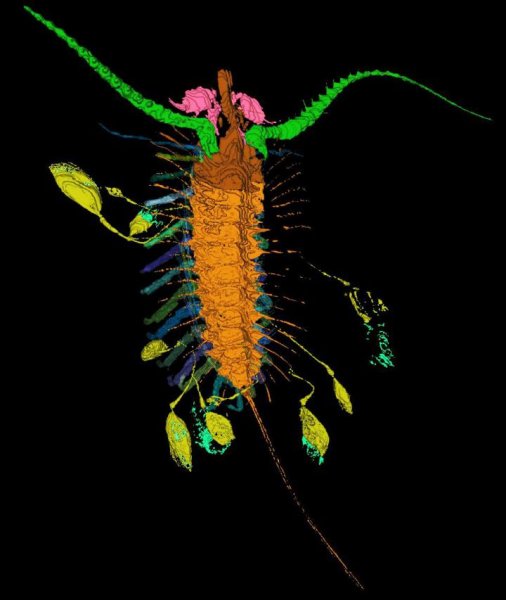
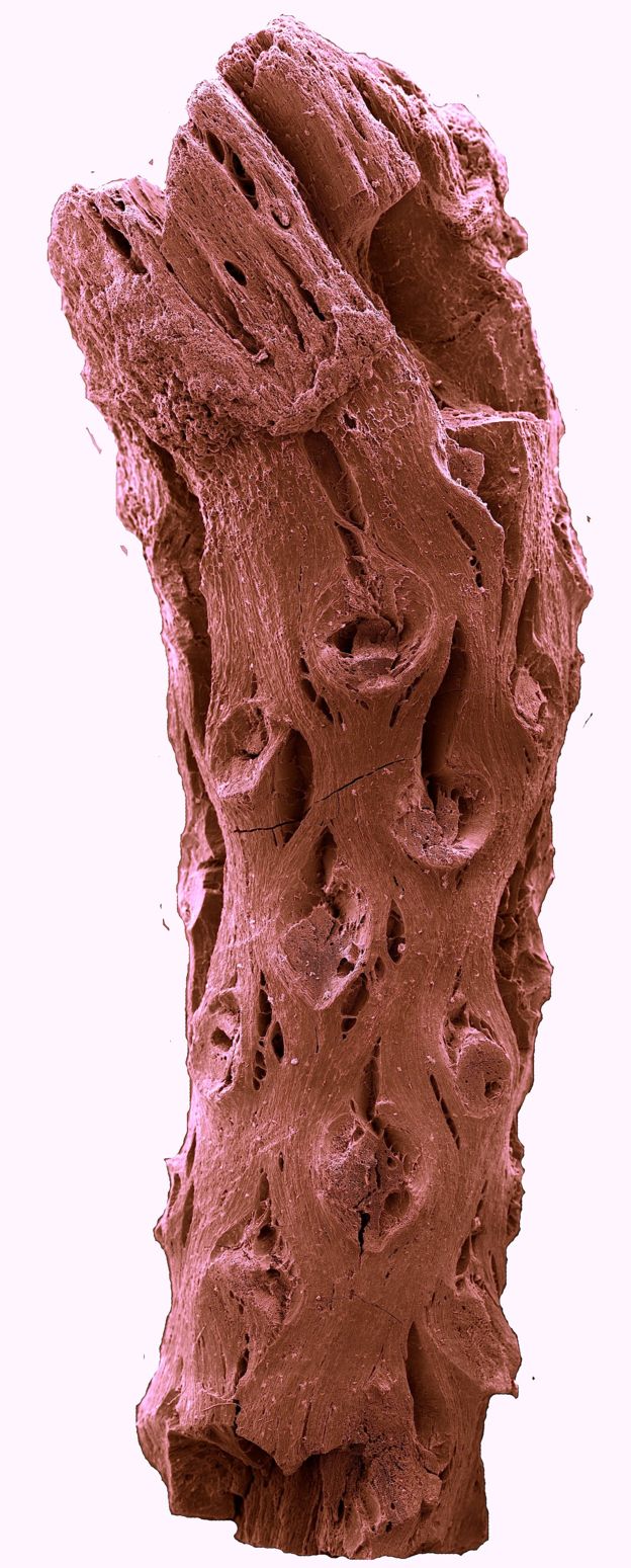
![Preserved Skin in the Fossil Colubrid Snake MNCN 66503 (A) Entire specimen; inset shows anterior. Cream-colored material is fossil skin. Numerals 1–7 indicate sample locations. (B) Overlapping scales. (C–E) Scanning electron micrographs (SEMs) of fractured vertical sections through the skin, showing epidermis (Epi), dermis (De), basement membrane (B), chromatophores (iridophores [I], melanophores [M], and xanthophores [X]), stratum spongiosum (Sp), stratum compactum (Sc), and collagen fibers (C). The voids in SEM images typically represent structures that have separated into the counterpart of the sample during preparation. (F–I) Details of iridophore (F), xanthophore (G), and melanophores (H and I). (J and K) Transmission electron micrographs of xanthophore (J) and melanophore (K).](http://www.worldfossilsociety.org/wp-content/uploads/2016/04/151209183454_1_540x360-1.jpg)
Preserved Skin in the Fossil Colubrid Snake MNCN 66503
(A) Entire specimen; inset shows anterior. Cream-colored material is fossil skin. Numerals 1–7 indicate sample locations.(B) Overlapping scales.(C–E) Scanning electron micrographs (SEMs) of fractured vertical sections through the skin, showing epidermis (Epi), dermis (De), basement membrane (B), chromatophores (iridophores [I], melanophores [M], and xanthophores [X]), stratum spongiosum (Sp), stratum compactum (Sc), and collagen fibers (C). The voids in SEM images typically represent structures that have separated into the counterpart of the sample during preparation.(F–I) Details of iridophore (F), xanthophore (G), and melanophores (H and I).(J and K) Transmission electron micrographs of xanthophore (J) and melanophore (K).
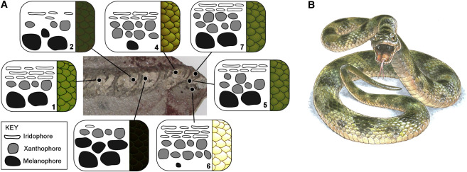
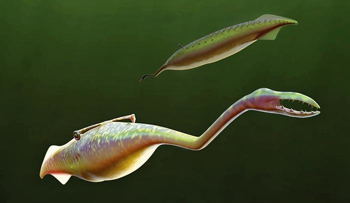
Color Reconstruction of the Fossil Snake MNCN 66503
(A) Schematic representation of the relative abundance and position of chromatophores in samples of skin from different body regions. Numerals denote samples discussed in the text. See also Tables S1 and S2.
(B) Color plate by Jim Robbins.
McNamara first came across the fossilized snake while conducting her PhD research on fossils from the Libros site in Spain, but she only recently analyzed the specimen. Her team discovered the mineralized skin cells when viewing the fossil under a high-powered scanning electron microscope and then matched the shapes up with pigment cells in modern snakes to determine what colors they might have produced.
“For the first time, we’re seeing that mineralized tissues can preserve evidence of color,” says McNamara. The researchers determined that the fossilized snakeskin had three types of pigment cells in various combinations: melanophores, which contain the pigment melanin; xanthophores, which contain carotenoid and pterin pigments; and iridophores, which create iridescence. All told, the snake was a mottled green and black, with a pale underside–colors that likely aided in daytime camouflage.
“Up until this discovery, the only prospect for skin color being preserved in fossils was organic remains related to melanin,” says McNamara. “But now that we know color can be preserved even for tissues that are mineralized, it’s very exciting.”
Calcium phosphate mainly shows up in fossil bones and shells, but records do exist of so-called phosphatized skin. This discovery opens the door for re-analysis of these fossils, occurring across a wide range of creatures and locations, for evidence of color preservation. And knowing the color of an animal can also clue researchers in to some aspects of its behavior and evolution.
“It’ll mean re-evaluating a lot of specimens that might have been overlooked,” says McNamara.
Citation :Cell Press. “A fossilized snake shows its true colors.” ScienceDaily. ScienceDaily, 31 March 2016. <www.sciencedaily.com/releases/2016/03/160331133402.htm
Key: WFS,Riffin T Sajeev,Russel T Sajeev,World Fossil Society


![Details of holotype (NIGP 163253) of Electrokoenenia yaksha Engel and Huang, gen. n. et sp. n., in mid-Cretaceous amber from northern Myanmar. a Frontal view of cheliceral chela as preserved (note that the right chela [left in image] is turned such that its leading edge is more angled toward the viewer, while the left is more in profile). b Reconstruction of chela, from observable details. c Detail of dorsal surface of prosoma and anterior legs (the fine lines radiating from body are not setae but rather minute, reflective fractures within the amber itself resulting from weakness around the inclusion)](http://www.worldfossilsociety.org/wp-content/uploads/2016/03/151209183454_1_540x360-2.gif)