Bandringa fossils revealed

Like salmon in reverse, long-snouted Bandringa sharks migrated downstream from freshwater swamps to a tropical coastline to spawn 310 million years ago, leaving behind fossil evidence of one of the earliest known shark nurseries.

That’s the surprising conclusion of University of Michigan paleontologist Lauren Sallan and a University of Chicago colleague, who reanalyzed all known specimens of Bandringa, a bottom-feeding predator that lived in an ancient river delta system that spanned what is today the Upper Midwest.

The new findings, scheduled for online publication Jan. 7 in the Journal of Vertebrate Paleontology, mark the earliest known example of shark migration — a behavior that persists today among species such as tiger sharks in Hawaii.

The Bandringa fossils, as reinterpreted by Sallan and Michael Coates, also reveal the only known example of a freshwater to saltwater shark migration, as well as the earliest example of a shark nursery where fossilized egg cases and juvenile sharks were preserved in the same sediments.

“This pushes migratory behavior in sharks way back,” said Sallan, an assistant professor in the U-M Department of Ecology and Evolutionary Biology. “These sharks bred in the open ocean and spent the rest of their lives in fresh water. No shark alive today is known to do that.”

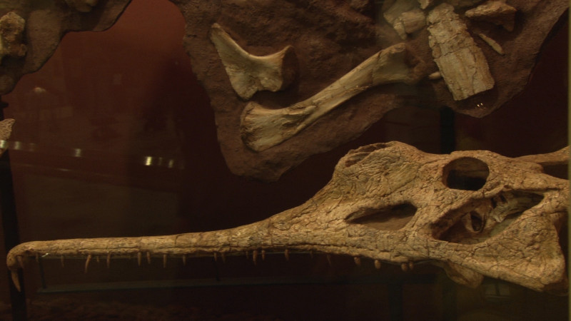
The long-extinct Bandringa is likely one of the earliest close relatives of modern sharks. It resembled present-day sawfish and paddlefish, with a spoon-billed snout up to half its body length. Juveniles were 4 to 6 inches long and grew into adults of up to 10 feet.

An artist's rendering of Bandringa, a 310 million-year-old shark originally found in fossil deposits from Mazon Creek, Illinois. University of Michigan paleontologist Lauren Sallan and a colleague say this bottom-feeding predator migrated to the ocean to spawn in shallow coastal waters and left behind fossil evidence of one of the earliest known shark nurseries.

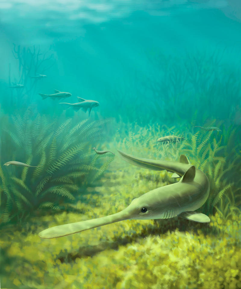
An artist’s rendering of Bandringa, a 310 million-year-old shark originally found in fossil deposits from Mazon Creek, Illinois. University of Michigan paleontologist Lauren Sallan and a colleague say this bottom-feeding predator migrated to the ocean to spawn in shallow coastal waters and left behind fossil evidence of one of the earliest known shark nurseries.

Bandringa was discovered in 1969 and soon became one of the most prized fossils from the well-known Mazon Creek deposits in northern Illinois. Until now, researchers believed that the genus Bandringa contained two species, one that lived in freshwater swamps and rivers and another that lived in the shallow ocean.

But after reevaluating fossils from 24 individuals, including latex “peels” of Bandringa’s scale-covered skin, Sallan and Coates concluded that Bandringa was a single species that lived, at various times during its life, in fresh, brackish and salt water.

The physical differences between the two purported species were due to different preservation processes at marine and freshwater locations, Sallan and Coates concluded. The freshwater sites tended to preserve bones and cartilage, while the marine sites preserved soft tissue.

By combining the complementary data sets from both types of fossil sites and reclassifying Bandringa as a single species, Sallan and Coates gained a far more complete picture of the extinct shark’s anatomy and discovered several previously unreported features. They include downward-directed jaws ideal for suction-feeding off the bottom, needle-like spines on the head and cheeks, and a complex array of sensory organs (electroreceptors and mechanoreceptors) on both the extended snout and body, suited for detecting prey in murky water.

Adult Bandringa sharks lived exclusively in freshwater swamps and rivers, according to Sallan and Coates. Females apparently traveled downstream to a tropical coastline to lay their eggs in shallow marine waters, a reverse version of the modern-day salmon’s sea-to-stream migration. At the time, the coastline of the super-continent Pangaea ran diagonally between the Mazon Creek freshwater and marine sites.

All the Bandringa fossils from the Mazon Creek marine sites are juveniles, and they were found alongside egg cases — protective capsules that enclose eggs of the next generation — belonging to an early species of shark. Adult Bandringa fossils have been found only at freshwater locations, including several in Ohio and Pennsylvania.

Sallan and Coates said that the juvenile Bandringa sharks hatched from the Mazon Creek egg cases, and that the deposit’s marine sites represent a shark nursery where females spawned and then departed, returning upstream to freshwater rivers and swamps.

“This is the first fossil evidence for a shark nursery that’s based on both egg cases and the babies themselves,” Sallan said. “It’s also the earliest evidence for segregation, meaning that juveniles and adults were living in different locations, which implies migration into and out of these nursery waters.”

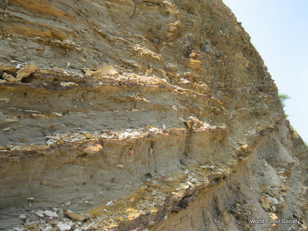
The Mazon Creek deposit is known for its extremely diverse, well-preserved fossils, with more than 320 animal species identified, according to the Illinois State Museum.

Many of those animals lived in shallow marine bays. Other plants and animals lived in swampy areas along rivers that emptied into the bay. When the remains of all these plants and animals sank to the bottom of the bay, they were rapidly buried by mud washing in from the river, which preserved them.

Financial support for the project was provided by the National Science Foundation, the University of Chicago, the University of Michigan and the Michigan Society of Fellows.

Role of rare earths in interpreting certain fossils

Until now, interpreting flattened fossils was a major challenge. Now, a new approach for the analysis of such fossils has been developed by a team bringing together researchers from the IPANEMA unit (CNRS / French Ministry of Culture and Communication), the Centre de Recherche sur la Paléobiodiversité et les Paléoenvironnements (CNRS / MNHN / UPMC) and the SOLEIL synchrotron. This non-destructive method makes use of chemical elements known as rare earths. By locating and quantifying such elements in trace amounts, it is possible to improve interpretation of fossil morphology. This enabled the researchers to describe not only the anatomy but also the environment of preservation of three fossils of Cretaceous age. Published on 29 January in the journal Plos One, the work should facilitate the interpretation of many flattened fossils, especially those that are exceptionally well conserved.

During the fossilization process, animal and plant remains are frequently flattened and compressed into two dimensions by the pressure of rocks, which sometimes proves to be a real obstacle to the study of such fossils. An added difficulty is that such flattened fossils undergo physical and chemical alteration during fossilization, making it even harder to interpret them. However, such fossils can contain invaluable information. In particular, when their anatomy is well preserved (in which case they are termed exceptionally well-preserved fossils), soft tissues such as muscles are also fossilized. However, locating such tissues remains particularly difficult due to the limited contrast attained in optical microscopy and to the limits of tomography[1], techniques that are today commonly used to study fossils.

Now, researchers from CNRS, the French Natural History Museum (MNHN) and the SOLEIL synchrotron have designed and developed a novel non-destructive approach based on the localization of rare earths. These chemical elements (yttrium and the lanthanides) are known to exist in fossils in trace amounts, typically from 1 to 1000 micrograms per gram of material. However, depending on the type of tissue, the quantity of trace elements incorporated during fossilization varies. Such preferential fixing makes it possible to distinguish between the anatomical parts of a fossil. This appears as a strong contrast between different chemical elements according to the types of fossil tissue, when the fossil is characterized using synchrotron fast X-ray fluorescence imaging[2]. To speed up the analysis, the team proposes a rapid method for differentiating tissue, based on the probabilistic nature of the data measured.

The scientists applied this approach to three fossils (two fishes and a shrimp) discovered in Morocco and dating from the Upper Cretaceous, around 100 million years ago. The contrasts revealed using this method enabled them to distinguish between hard tissues (bones and shells) and soft tissues (muscles and other fossilized organs). In particular, this enabled them to discover the previously hidden anatomical features of a fossil fish of which only one specimen is known, one of whose skull bones is in the form of a wide, notched blade.

This new approach makes it possible to obtain a detailed, accurate view of the anatomy of a fossil without damaging it and without the need for prior delicate sample preparation. It is particularly suitable for flattened fossils given that X-rays penetrate the fossil to a depth of a few fractions of a millimeter. The technique also revealed certain bones concealed under a fine layer of rock, enabling them to be viewed directly. For instance, it enabled certain concealed appendages of a fossil shrimp to be viewed, such as the legs and the antennae, which hold important information for the study of its relationship to other shrimps. In addition, rare earth content reflects the environment in which a fossil is preserved, such as connectivity to surrounding water networks, local physico-chemical conditions, and the properties of the mineral phases making up the fossils, thus enabling them to be better described.

The work should therefore facilitate the interpretation of flattened fossils, which are very common in the fossil record. It opens up new prospects not only for paleoenvironmental studies but also for a better understanding of long-term fossilization processes.

The research was carried out as part of the IPANEMA research platform, inaugurated on 12 September 2013 by Geneviève Fioraso, French Minister of Research and Higher Education. IPANEMA is a joint unit of CNRS and the French Ministry of Culture and Communication, set up in partnership with the French National History Museum, with support from the SOLEIL synchrotron which hosts it, and the European Commission (FP7 CHARISMA project).

[1] Tomography is a technique based on the reconstruction of virtual cross-sections of a 3-dimensional object from a large number of X-ray exposures.

[2] X-ray fluorescence is the secondary X-ray emission from atoms bombarded with X-rays. The emission spectrum is characteristic of the chemical elements making up the sample. Used in imaging mode, it can be used to locate these elements. In this study, the very high intensity of the synchrotron radiation makes it possible to access elements present in trace amounts, which is unachievable with laboratory X-rays.

Bones of a previously unknown species prove to be one of the oldest seabirds

Fossils discovered in Canterbury, New Zealand reveal the nature of one of the world’s oldest flying seabirds. Thought to have lived between 60.5 and 61.6 million years ago, the fossil is suggested to have formed shortly after the extinction of dinosaurs and many marine organisms.

Bones of the bird were discovered in 2009 by Leigh Love, an amateur fossil collector. The new species, Australornis lovei has been named as such in honour of Love’s discovery.

The bird lacks key morphological features of penguins, though it was found near the fossils of the Waimanu manneringi, the oldest penguin, of which it is also estimated to be the same age.

The research is published in Journal of the Royal Society of New Zealand by Dr Gerald Mayr and Dr Paul Scofield. The authors claim the discovery ‘represents one of the most significant records of a marine Paleocene bird from the Southern Hemisphere’ and supports the ’emerging view that most modern birds were already diversified in the earliest Paleogene’.

Despite the distinctness of this new species, its derived features are not limited to a single bird group. It does resemble an extinct species from Antarctica, however, highlighting the links between Antarctica and New Zealand in the late Cretaceous period.

Courtesy: ScienceDaily

Using rare earths to interpret certain fossils

Until now, interpreting flattened fossils was a major challenge. Now, a new approach for the analysis of such fossils has been developed by a team bringing together researchers from the IPANEMA unit (CNRS / French Ministry of Culture and Communication), the Centre de Recherche sur la Paléobiodiversité et les Paléoenvironnements (CNRS / MNHN / UPMC) and the SOLEIL synchrotron. This non-destructive method makes use of chemical elements known as rare earths. By locating and quantifying such elements in trace amounts, it is possible to improve interpretation of fossil morphology. This enabled the researchers to describe not only the anatomy but also the environment of preservation of three fossils of Cretaceous age. Published on 29 January in the journal Plos One, the work should facilitate the interpretation of many flattened fossils, especially those that are exceptionally well conserved. Share 12 During the fossilization process, animal and plant remains are frequently flattened and compressed into two dimensions by the pressure of rocks, which sometimes proves to be a real obstacle to the study of such fossils. An added difficulty is that such flattened fossils undergo physical and chemical alteration during fossilization, making it even harder to interpret them. However, such fossils can contain invaluable information. In particular, when their anatomy is well preserved (in which case they are termed exceptionally well-preserved fossils), soft tissues such as muscles are also fossilized. However, locating such tissues remains particularly difficult due to the limited contrast attained in optical microscopy and to the limits of tomography[1], techniques that are today commonly used to study fossils. Now, researchers from CNRS, the French Natural History Museum (MNHN) and the SOLEIL synchrotron have designed and developed a novel non-destructive approach based on the localization of rare earths. These chemical elements (yttrium and the lanthanides) are known to exist in fossils in trace amounts, typically from 1 to 1000 micrograms per gram of material. However, depending on the type of tissue, the quantity of trace elements incorporated during fossilization varies. Such preferential fixing makes it possible to distinguish between the anatomical parts of a fossil. This appears as a strong contrast between different chemical elements according to the types of fossil tissue, when the fossil is characterized using synchrotron fast X-ray fluorescence imaging[2]. To speed up the analysis, the team proposes a rapid method for differentiating tissue, based on the probabilistic nature of the data measured. The scientists applied this approach to three fossils (two fishes and a shrimp) discovered in Morocco and dating from the Upper Cretaceous, around 100 million years ago. The contrasts revealed using this method enabled them to distinguish between hard tissues (bones and shells) and soft tissues (muscles and other fossilized organs). In particular, this enabled them to discover the previously hidden anatomical features of a fossil fish of which only one specimen is known, one of whose skull bones is in the form of a wide, notched blade. This new approach makes it possible to obtain a detailed, accurate view of the anatomy of a fossil without damaging it and without the need for prior delicate sample preparation. It is particularly suitable for flattened fossils given that X-rays penetrate the fossil to a depth of a few fractions of a millimeter. The technique also revealed certain bones concealed under a fine layer of rock, enabling them to be viewed directly. For instance, it enabled certain concealed appendages of a fossil shrimp to be viewed, such as the legs and the antennae, which hold important information for the study of its relationship to other shrimps. In addition, rare earth content reflects the environment in which a fossil is preserved, such as connectivity to surrounding water networks, local physico-chemical conditions, and the properties of the mineral phases making up the fossils, thus enabling them to be better described. The work should therefore facilitate the interpretation of flattened fossils, which are very common in the fossil record. It opens up new prospects not only for paleoenvironmental studies but also for a better understanding of long-term fossilization processes. The research was carried out as part of the IPANEMA research platform, inaugurated on 12 September 2013 by Geneviève Fioraso, French Minister of Research and Higher Education. IPANEMA is a joint unit of CNRS and the French Ministry of Culture and Communication, set up in partnership with the French National History Museum, with support from the SOLEIL synchrotron which hosts it, and the European Commission (FP7 CHARISMA project). [1] Tomography is a technique based on the reconstruction of virtual cross-sections of a 3-dimensional object from a large number of X-ray exposures. [2] X-ray fluorescence is the secondary X-ray emission from atoms bombarded with X-rays. The emission spectrum is characteristic of the chemical elements making up the sample. Used in imaging mode, it can be used to locate these elements. In this study, the very high intensity of the synchrotron radiation makes it possible to access elements present in trace amounts, which is unachievable with laboratory X-rays.

Courtesy – ScienceDaily

New Mid-Cretaceous (Latest Albian) Dinosaurs from Winton, Queensland, Australia

Background

Australia’s dinosaurian fossil record is exceptionally poor compared to that of other similar-sized continents. Most taxa are known from fragmentary isolated remains with uncertain taxonomic and phylogenetic placement. A better understanding of the Australian dinosaurian record is crucial to understanding the global palaeobiogeography of dinosaurian groups, including groups previously considered to have had Gondwanan origins, such as the titanosaurs and carcharodontosaurids.

Methodology/Principal Findings

We describe three new dinosaurs from the late Early Cretaceous (latest Albian) Winton Formation of eastern Australia, including; Wintonotitan wattsi gen. et sp. nov., a basal titanosauriform; Diamantinasaurus matildae gen. et sp. nov., a derived lithostrotian titanosaur; and Australovenator wintonensis gen. et sp. nov., an allosauroid. We compare an isolated astragalus from the Early Cretaceous of southern Australia; formerly identified as Allosaurus sp., and conclude that it most-likely represents Australovenator sp.

Conclusion/Significance

The occurrence of Australovenator from the Aptian to latest Albian confirms the presence in Australia of allosauroids basal to the Carcharodontosauridae. These new taxa, along with the fragmentary remains of other taxa, indicate a diverse Early Cretaceous sauropod and theropod fauna in Australia, including plesiomorphic forms (e.g. Wintonotitan and Australovenator) and more derived forms (e.g. Diamantinasaurus).

Silhouettes of the three new dinosaurs showing the material currently known from their respective holotypes. show more  A–B. Diamantinasaurus matildae gen. et sp. nov. (AODF 603); A. Right side, B. Left side. C. Wintonotitan wattsi gen. et sp. nov. (QMF 7292); Left view. D. Australovenator wintonensis gen. et sp. nov. (QMF 7292); Right view. Artwork: T. Tischler, Australian Age of Dinosaurs Museum of Natural History.  doi:10.1371/journal.pone.0006190.g002

Silhouettes of the three new dinosaurs showing the material currently known from their respective holotypes.
A–B. Diamantinasaurus matildae gen. et sp. nov. (AODF 603); A. Right side, B. Left side. C. Wintonotitan wattsi gen. et sp. nov. (QMF 7292); Left view. D. Australovenator wintonensis gen. et sp. nov. (QMF 7292); Right view. Artwork: T. Tischler, Australian Age of Dinosaurs Museum of Natural History.
doi:10.1371/journal.pone.0006190.g002

Dorsal ribs of Diamantinasaurus matildae. show more  Mid-dorsal rib in lateral (A), anterior (B) and posterior (C) views. Posterior dorsal rib in posterior (A) and anterior (B) views.  doi:10.1371/journal.pone.0006190.g003

Dorsal ribs of Diamantinasaurus matildae.
Mid-dorsal rib in lateral (A), anterior (B) and posterior (C) views. Posterior dorsal rib in posterior (A) and anterior (B) views.
doi:10.1371/journal.pone.0006190.g003

Scapula and sternal plate of Diamantinasaurus matildae. show more  Right scapula in lateral view (A). Left sternal plate in ventral view (B). Abbreviations: ac, acromial blade; crsp, cranial ridge of sternal plate; cs, coracoid suture; sb, scapular blade; sg, scapula glenoid. Dashed line indicates suggested area missing from specimen.  doi:10.1371/journal.pone.0006190.g004

Scapula and sternal plate of Diamantinasaurus matildae.
Right scapula in lateral view (A). Left sternal plate in ventral view (B). Abbreviations: ac, acromial blade; crsp, cranial ridge of sternal plate; cs, coracoid suture; sb, scapular blade; sg, scapula glenoid. Dashed line indicates suggested area missing from specimen.
doi:10.1371/journal.pone.0006190.g004


For Article details and more pictures:

Citation: Hocknull SA, White MA, Tischler TR, Cook AG, Calleja ND, et al. (2009) New Mid-Cretaceous (Latest Albian) Dinosaurs from Winton, Queensland, Australia. PLoS ONE 4(7): e6190. doi:10.1371/journal.pone.0006190

Editor: Paul Sereno, University of Chicago, United States of America


“Yongjinglong datangi” The New dinosaur fossil discovered in China

Scientists have discovered the fossil of a 60-feet long plant-eating dinosaur in China that lived about 100 million years ago.

A team led by University of Pennsylvania paleontologists has characterised the new dinosaur based on fossil remains found in northwestern China.

The species, a plant-eating sauropod named Yongjinglong datangi, roamed during the Early Cretaceous period, more than 100 million years ago.

This sauropod belonged to a group known as Titanosauria, members of which were among the largest living creatures to ever walk the Earth.

At roughly 50-60 feet long, the Yongjinglong individual discovered was a medium-sized Titanosaur. Anatomical evidence, however, points to it being a juvenile; adults may have been larger.

The finding, reported in the journal PLOS ONE, helps clarify relationships among several sauropod species that have been found in the last few decades in China and elsewhere.

An undated image released by the Royal Belgian Institute of Natural Sciences shows the skeleton of a dinosaur dubbed Aurornis xui that roamed China during the middle to late Jurassic period. File photo

An undated image released by the Royal Belgian Institute of Natural Sciences shows the skeleton of a dinosaur dubbed Aurornis xui that roamed China during the middle to late Jurassic period. File photo

Its features suggest that Yongjinglong is among the most derived, or evolutionarily advanced, of the Titanosaurs yet discovered from Asia.

Until very recently, the U.S. was the epicentre for dinosaur diversity, but China surpassed the U.S. in 2007 in terms of species found, researchers said.

This latest discovery was made in the southeastern Lanzhou-Minhe Basin of China’s Gansu Province.

The remains consisted of three teeth, eight vertebrae, the left shoulder blade, and the right radius and ulna.

The anatomical features of the bones bear some resemblance to another Titanosaur that had been discovered in China in 1929, named Euhelopus zdanskyi.

“The shoulder blade was very long, nearly 2 meters, with sides that were nearly parallel, unlike many other Titanosaurs whose scapulae bow outward,” Doctoral student Liguo Li said.

The scapula was so long that it did not appear to fit in the animal’s body cavity if placed in a horizontal or vertical orientation, as is the case with other dinosaurs.

source “The Hindu”

Rare female phytosaur skull found in West Texas more than 200 million years old

In the dangerous waters of an ancient oxbow lake created by a flooded and unnamed meandering river, the female phytosaur died and sank to the bottom 205 million years ago. About 40 yards away the remains of a larger male also came to rest, and both disappeared in a tomb of soil and sediment.

Evidence for the cause of their deaths and the rest of their bodies have vanished with time, but their skulls remained. After careful research, a Texas Tech paleontologist says he and others have discovered a new species of the Triassic-age monster in the wilds of West Texas.

Their findings were published in the peer-reviewed journal Earth and Environmental Science Transactions of the Royal Society of Edinburgh.

The female skull still had its teeth intact, which made the find even rarer. Credit: Photo by Lacey Nobles

The female skull still had its teeth intact, which made the find even rarer.
Credit: Photo by Lacey Nobles

Bill Mueller, assistant curator of Paleontology at the Museum of Texas Tech University, said the team named their find Machaeroprosopus lottorum after the Lott family who own the ranch on which the animal was discovered.

“We found them in an area we’d been excavating in,” Mueller said. “I think we’ve gotten four skulls out of that area already. Doug Cunningham found this specimen, and then we dug it up. When he found it, just the very back end of the skull was sticking out of the ground. The rest was buried. We excavated it and brought it into the museum to finish preparation.”

Cunningham, currently a field research assistant at the museum and a retired firefighter, remembered finding the unusual female skull on June 27, 2001. After removing it from the mudstone, he recalls looking it over carefully with others and wondering if his discovery would add a new animal to science.

“It was really well preserved with the teeth and everything,” Cunningham said. “Finding one with teeth is pretty rare. It was so odd, but when they come out of the ground, you have a long way to go to actually see what you have because they’re still covered in matrix. We were all kind of in awe of it. It had this long, skinny snout. It was quite a bit different. It took me years to get it prepped and ready. At the time, I was working full-time and I did that on my days off.” By looking an opening on the skull called the supratemporal fenestra, the snout and the shape of the bones at the back of the head, the team compared it to other phytosaurs and determined they’d discovered a separate species.

While West Texas is dry and dusty today, Mueller said the landscape looked more like a swampy, tropical rainforest during the Triassic period. Our planet’s landmasses had converged to form the supercontinent of Pangaea. In the forest undergrowth covered by tall conifers and choked with ferns, phytosaurs lurked beneath the water and waited for prey.

“A phytosaur resembles a crocodile,” Mueller said. “They had basically the same lifestyle as the modern crocodile by living in and around the water, eating fish, and whatever animals came to the margins of the rivers and lakes. But one of the big differences is the external nares, the nose, is back up next to its eyes instead of at the end of its snout.”

Mueller said scientists can tell the sexes of the animals by a distinctive feature on males. A bony crest stretched from the nostrils by the eyes to the tip of the animal’s beak — a feature lady phytosaurs probably found sexy.

Judging by the female’s skull size, which is more than three feet in length, Mueller guessed she would have measured 16 to 17 feet in length from nose to tail tip. The male would have measured about 17 to 18 feet. Their thin jaws suggested they hunted mainly fish as opposed to big prey.

Mueller said phytosaurs lived throughout the Triassic period from 230 to 203 million years ago, but died out during a mysterious mass extinction. Highly successful animals, they are commonly found because these animals liked to live in swampy areas and were more likely to become covered in sediment and fossilized.

Dinosaur-chewing mammals leave behind oldest known tooth marks

Paleontologists have discovered the oldest mammalian tooth marks yet on the bones of ancient animals, including several large dinosaurs. They report their findings in a paper published online June 16 in the journal Paleontology.

A close-up of the tooth marks gouged in the rib bone of a large dinosaur by a mammal that lived 75 million years ago.

A close-up of the tooth marks gouged in the rib bone of a large dinosaur by a mammal that lived 75 million years ago.

Nicholas Longrich of Yale University and Michael J. Ryan of the Cleveland Museum of Natural History came across several of the bones while studying the collections of the University of Alberta Laboratory for Vertebrate Palaeontology and the Royal Tyrrell Museum of Palaeontology. They also found additional bones displaying tooth marks during fieldwork in Alberta, Canada. The bones are all from the Late Cretaceous epoch and date back about 75 million years.

The pair discovered tooth marks on a femur bone from a Champsosaurus, an aquatic reptile that grew up to five feet long; the rib of a dinosaur, most likely a hadrosaurid or ceratopsid; the femur of another large dinosaur that was likely an ornithischian; and a lower jaw bone from a small marsupial.

The researchers believe the marks were made by mammals because they were created by opposing pairs of teeth — a trait seen only in mammals from that time. They think they were most likely made by multituberculates, an extinct order of archaic mammals that resemble rodents and had paired upper and lower incisors. Several of the bones display multiple, overlapping bites made along the curve of the bone, revealing a pattern similar to the way people eat corn on the cob.

The animals that made the marks were about the size of a squirrel and were most likely gnawing on the bare bones for minerals rather than for meat, said Longrich. “The bones were kind of a nutritional supplement for these animals.”

There are likely many other instances of mammalian tooth marks on other bones that have yet to be identified, including older examples, said Longrich. “The marks stood out for me because I remember seeing the gnaw marks on the antlers of a deer my father brought home when I was young,” he said. “So when I saw it in the fossils, it was something I paid attention to.”

But he points out that the Late Cretaceous creatures that chewed on these bones were not nearly as adept at gnawing as today’s rodents, which developed that ability long after dinosaurs went extinct.

Courtesy: Yale University. “Dinosaur-chewing mammals leave behind oldest known tooth marks.” ScienceDaily. ScienceDaily, 17 June 2010. <www.sciencedaily.com/releases/2010/06/100616161207.htm>.

Old Bird, New World: Did the South American Hoatzins Originate in Europe?

The oldest fossil discoveries from France show that hoatzins once existed in Europe.

Where did hoatzins come from? These unusual birds, only one species of which exists in South America today, originated in the Old World. Studies of the oldest known fossils of Hoatzin ancestors have now shown that these birds existed around 34 million years ago in Europe. Paleornithologists at the Senckenberg Gesellschaft für Naturforschung and Flinders University in Adelaide published their findings this week in the specialist journal Naturwissenschaften.

The Hoatzin (Opisthocomus hoazin) is also known as the Stinkbird or Canje Pheasant and today exists only in parts of South America. Its relationship among birds is as unclear as its evolutionary history. Until recently, South America was considered to be the area of origin of these birds. Then, however, fossils from Africa were described, and new discoveries from Africa and Europe now prove conclusively that hoatzins reached South America from the Old World. A study published in January 2014 in the ornithological journal “The Auk” provided the very first evidence of largely modern Hoatzins from the Miocene (15 million years ago) in Africa.

Protoazin parisiensis and Opisthocomus hoazin. (Credit: © Senckenberg)

Protoazin parisiensis and Opisthocomus hoazin. (Credit: © Senckenberg)

The oldest fossils attributed to hoatzins were found more than 100 years ago near Paris. But only now have paleornithologists been able to show that these remains belong to ancestors of the modern Hoatzin. The fossils belong to a newly described species, Protoazin parisiensis (“proto-Hoatzin from Paris”). The re-interpretation of these bones indicates that hoatzins lived in Europe as early as the late Eocene, i.e. around 34 million years ago.

“This supports the theory that hoatzins originated in the Old World,” says Dr Gerald Mayr of the Senckenberg Research Institute in Frankfurt. In the opinion of Dr Vanesa De Pietri of Flinders University in Australia, it is “a further impressive example that the South American avian fauna contains numerous relicts that were once much more widespread.”

Most notably, Hoatzins appear to have become extinct in Europe much earlier than in Africa, where the latest fossils were dated as of Miocene age (15 million years ago) and are thus only around half as old. The disappearance of these birds might be connected to a period when numerous new animal species migrated from Asia to Europe during the so-called “Grande Coupure” around 34 million years ago. These included tree-dwelling carnivorous mammals who may have posed a threat to hoatzin nestlings, which are raised in open nests. Because hoatzins can fly short distances only, the adult birds are also easy prey. In Africa, by contrast, similar tree-dwelling carnivorous mammals are shown to have existed much later.

Digestion specialist and climbing artist

The present-day Hoatzin exhibits a special mode of digestion. These herbivores pre-digest their food in this crop before further processing in the stomach and intestines. Similar to the rumen of a cow — a digestive knack that has not been mastered by any other bird. Skeletal features show that Old World hoatzins already had a large crop. Another special feature of the Hoatzin are the claws on the wings of the chicks, which enable the hatchlings to climb trees.

Ancient marine fossil found

A unique fossil found at the tip of the South Island has been recognised as a 20-million-year-old marine ancestor.

The fossil, discovered near Cape Farewell, was collected and analysed by geologists at the University of Otago.

They described it as a close relation to the ancestors of modern dolphins and toothed whales.

Professor Ewan Fordyce has dubbed the fossil Papahu taitapu.

Professor Ewan Fordyce has dubbed the fossil Papahu taitapu.

Professor Ewan Fordyce and his PhD student, Dr Gabriel Aguirre, dubbed the fossil Papahu taitapu, and said it was the first of its kind ever found. This made it a significant addition to the fossil record of dolphins from the southern hemisphere.

Papahu is estimated to have lived between 19 million and 20 million years ago, making it one of the few dolphins dating to the start of the Miocene epoch.

“New Zealand was substantially under water back then,” Fordyce said.

“There were wide shallow seaways. We know also that the climate was fairly warm, a lot warmer than today, with water temperatures down south about 18 degrees [Celsius] or warmer – good for swimming!”

Judging from the size of Papahu’s skull, the dolphin was about 2 metres long – roughly the size of a common dolphin – and likely had “many simple conical teeth”, he said.

Features of the skull showed that it was distinct from all previously reported fossils, which was why the dolphin could be formally named as a new form.

“When we compared Papahu with both modern and fossil dolphins we found that it belongs in a diverse and structurally variable group of ancient dolphins that evolved and spread worldwide 19 million to 35 million years ago,” Fordyce said.

“All of those ancient dolphins including Papahu and others, such as shark-toothed dolphins, are now extinct.

“They have been replaced by the modern dolphins and toothed whales, which diversified within the last 19 million years.”

It was not clear why Papahu and other ancient dolphins went extinct.

source: © Fairfax NZ News