

 April 5th, 2013
  Riffin
 It has been assumed that the unusual tail club of ankylosaurid dinosaurs was used actively as a weapon, but the biological feasibility of this behaviour has not been examined in detail. Ankylosaurid tail clubs are composed of interlocking vertebrae, which form the handle, and large terminal osteoderms, which form the knob.
Methodology/Principal Findings
Computed tomographic (CT) scans of several ankylosaurid tail clubs referred to Dyoplosaurus and Euoplocephalus, combined with measurements of free caudal vertebrae, provide information used to estimate the impact force of tail clubs of various sizes. Ankylosaurid tails are modeled as a series of segments for which mass, muscle cross-sectional area, torque, and angular acceleration are calculated. Free caudal vertebrae segments had limited vertical flexibility, but the tail could have swung through approximately 100° laterally. Muscle scars on the pelvis record the presence of a large M. longissimus caudae, and ossified tendons alongside the handle represent M. spinalis. CT scans showed that knob osteoderms were predominantly cancellous, which would have lowered the rotational inertia of the tail club and made it easier to wield as a weapon.
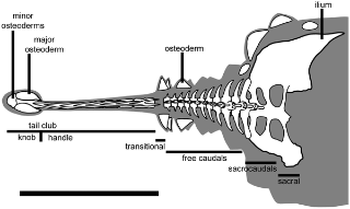
Ankylosaurid tail reconstructed from ROM 784; ROM 784 lacks the transitional caudal vertebra and the anterior portion of the pelvis. Scale bar equals 1 m. Modified from Arbour et al. (in press).
doi:10.1371/journal.pone.0006738.g001
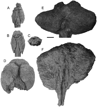
Morphology of ankylosaurid tail clubs.
A) UALVP 47273, dorsal view. B) ROM 784 dorsal view and C) posterior view, D) UALVP 16247 dorsal view, E) AMNH 5245 dorsal view, and F) ROM 788 ventral view.
doi:10.1371/journal.pone.0006738.g002
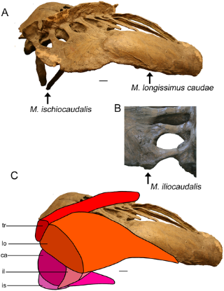
Origins of tail muscles on the pelvis.
A) AMNH 5409 (Euoplocephalus) pelvis, posterior right dorsolateral view. M. ischiocaudalis originates at the distal terminus of the ischium. The origin of M. longissimus caudae is marked by a long, pronounced ridge and rugose area on the lateral aspect of the ilium. The posterior terminus of the ilium is partially reconstructed. B) AMNH 5337 (Euoplocephalus) pelvis, dorsal view, anterior up, showing the posterior terminus of the left ilium. M. iliocaudalis originates from a large knob. C) AMNH 5409, same view as (A), with reconstructed musculature. The muscles are cut posteriorly to show their relationships in cross-section. M. caudofemoralis longus originates on the transverse processes of the free caudal vertebrae, and inserts on the fourth trochanter of the femur (not shown). M. transversospinalis originates and inserts on the neural spines. Scale bars equal 10 cm. Abbreviations are as follows: ca = M. caudofemoralis longus, il = M. iliocaudalis, is = M. ischiocaudalis, lo = M. longissimus caudae, tr = M. transversospinalis.
doi:10.1371/journal.pone.0006738.g008
Conclusions/Significance
Large knobs could generate sufficient force to break bone during impacts, but average and small knobs could not. Tail swinging behaviour is feasible in ankylosaurids, but it remains unknown whether the tail was used for interspecific defense, intraspecific combat, or both.
Citation: Arbour VM (2009) Estimating Impact Forces of Tail Club Strikes by Ankylosaurid Dinosaurs. PLoS ONE 4(8): e6738. doi:10.1371/journal.pone.0006738
Editor: Andrew Allen Farke, Raymond M. Alf Museum of Paleontology, United States of America
 Posted in
  Tags:
Tags: 