

 September 4th, 2013
  Riffin
 The pectoral and pelvic girdles support paired fins and limbs, and have transformed significantly in the diversification of gnathostomes or jawed vertebrates (including osteichthyans, chondrichthyans, acanthodians and placoderms). For instance, changes in the pectoral and pelvic girdles accompanied the transition of fins to limbs as some osteichthyans (a clade that contains the vast majority of vertebrates – bony fishes and tetrapods) ventured from aquatic to terrestrial environments. The fossil record shows that the pectoral girdles of early osteichthyans (e.g., Lophosteus, Andreolepis, Psarolepis and Guiyu) retained part of the primitive gnathostome pectoral girdle condition with spines and/or other dermal components. However, very little is known about the condition of the pelvic girdle in the earliest osteichthyans. Living osteichthyans, like chondrichthyans (cartilaginous fishes), have exclusively endoskeletal pelvic girdles, while dermal pelvic girdle components (plates and/or spines) have so far been found only in some extinct placoderms and acanthodians. Consequently, whether the pectoral and pelvic girdles are primitively similar in osteichthyans cannot be adequately evaluated, and phylogeny-based inferences regarding the primitive pelvic girdle condition in osteichthyans cannot be tested against available fossil evidence.
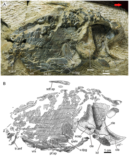
Figure 1. Guiyu oneiros Zhu et al., 2009.
A. New articulated specimen of Guiyu oneiros (V17914, lateral view) from the Kuanti Formation (Late Ludlow, Silurian), Qujing, Yunnan, showing a right dermal pelvic girdle in near-natural position. Red arrow points to the anterior end of the fish. B. Interpretative drawing. Abbreviations: ba.sc, basal scales of pelvic fin; cla, clavicle; cle, cleithrum; icl, interclavicle; ipelv, interpelvic plate; pelv.sp, pelvic fin spine; scap, scapulocoracoid; sdf.sp, second dorsal fin spine; tr.anf, lepidotrichia of anal fin; v.dpg, ventral lamina of dermal pelvic girdle; vrs, ventral ridge scale.
doi:10.1371/journal.pone.0035103.g001
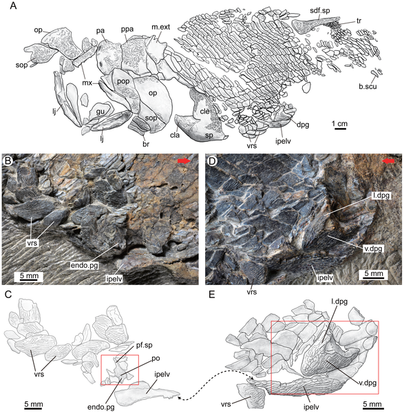
Figure 2. The holotype (V15541) of Guiyu oneiros Zhu et al., 2009.
A. Interpretative drawing of the part to show the position of the newly identified left pelvic girdle with dermal and endoskeletal components. B–C. Close-up of the counterpart to show the endoskeletal pelvic girdle in internal view (B) and interpretative drawing (C). D–E. Close-up of the part to show the dermal pelvic girdle in lateral view (D) and interpretative drawing (E). Red arrows point to the anterior end of the fish. The red rectangles indicate the close-up areas in Figure 3A and Figure 3B. The double arrows point to the corresponding positions of the fractured interpelvic plate in part (E) and counterpart (C). Abbreviations: br, branchiostegal ray; b.scu, basal scute; cla, clavicle; cle, cleithrum; dpg, dermal pelvic girdle; endo.pg, endoskeletal pelvic girdle; gu, gular; ipelv, interpelvic plate; l.dpg, lateral lamina of dermal pelvic girdle; lj, lower jaw; m.ext, median extrascapular; mx, maxillary; op, opercular; pa, parietal shield; pf.sp, pelvic fin spine; po, foramina for pterygial nerves and vessels; pop, preopercular; ppa, postparietal shield; sdf.sp, second dorsal fin spine; sop, subopercular; sp, pectoral fin spine; tr, lepidotrichia; v.dpg, ventral lamina of dermal pelvic girdle; vrs, ventral ridge scale.
doi:10.1371/journal.pone.0035103.g002
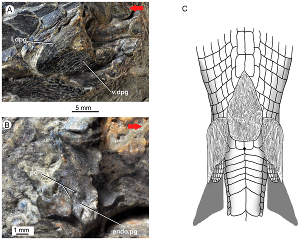
Figure 3. Guiyu oneiros Zhu et al., 2009.
A. Close-up of the holotype (in part) to show the dermal pelvic girdle in lateral view. B. Close-up of the holotype (in counterpart) to show the endoskeletal pelvic girdle in internal view. C. Tentative life restoration in ventral view to show the paired pelvic girdles and unpaired interpelvic plate. Red arrows point to the anterior end of the fish. Abbreviations: endo.pg, endoskeletal pelvic girdle; l.dpg, lateral lamina of dermal pelvic girdle; po, foramina for pterygial nerves and vessels; v.dpg, ventral lamina of dermal pelvic girdle.
doi:10.1371/journal.pone.0035103.g003
![Figure 4. Guiyu oneiros Zhu et al., 2009. show more A. Close-up of the three median dorsal plates (Md1-Md3) of the holotype V15541. B–C. A disarticulated third median dorsal plate (Md3) bearing the first dorsal fin spine in external (B) and internal (C) views, V17914.2. D. A disarticulated third median dorsal plate (Md3) in external view, V17914.3. E. Revised restoration of Guiyu oneiros in lateral view, based on [24] for the cranial portion, and new data in this work. Abbreviations: Md1–Md3, first to third median dorsal plates, with the third bearing the first dorsal fin spine and the endoskelatal basal plate. doi:10.1371/journal.pone.0035103.g004](http://worldfossilsociety.org/wp-content/uploads/2013/09/121219174150-large4.png)
Figure 4. Guiyu oneiros Zhu et al., 2009.
A. Close-up of the three median dorsal plates (Md1-Md3) of the holotype V15541. B–C. A disarticulated third median dorsal plate (Md3) bearing the first dorsal fin spine in external (B) and internal (C) views, V17914.2. D. A disarticulated third median dorsal plate (Md3) in external view, V17914.3. E. Revised restoration of Guiyu oneiros in lateral view, based on [24] for the cranial portion, and new data in this work. Abbreviations: Md1–Md3, first to third median dorsal plates, with the third bearing the first dorsal fin spine and the endoskelatal basal plate.
doi:10.1371/journal.pone.0035103.g004
Methodology/Principal Findings
Here we report the first discovery of spine-bearing dermal pelvic girdles in early osteichthyans, based on a new articulated specimen of Guiyu oneiros from the Late Ludlow (Silurian) Kuanti Formation, Yunnan, as well as a re-examination of the previously described holotype. We also describe disarticulated pelvic girdles of Psarolepis romeri from the Lochkovian (Early Devonian) Xitun Formation, Yunnan, which resemble the previously reported pectoral girdles in having integrated dermal and endoskeletal components with polybasal fin articulation.
Conclusions/Significance
The new findings reveal hitherto unknown similarity in pectoral and pelvic girdles among early osteichthyans, and provide critical information for studying the evolution of pelvic girdles in osteichthyans and other gnathostomes.
Citation: Zhu M, Yu X, Choo B, Qu Q, Jia L, et al. (2012) Fossil Fishes from China Provide First Evidence of Dermal Pelvic Girdles in Osteichthyans. PLoS ONE 7(4): e35103. doi:10.1371/journal.pone.0035103
Editor: Ulrich Joger, State Natural History Museum, Germany
 Posted in
  Tags:
Tags: 