

 July 17th, 2021
  Riffin
 @WFS,World Fossil Society,Riffin T Sajeev,Russel T Sajeev
Evidence of preserved collagen in an Early Jurassic sauropodomorph dinosaur revealed by synchrotron FTIR microspectroscopy
Lee, YC., Chiang, CC., Huang, PY. et al.
Nat Commun 8, 14220 (2017). https://doi.org/10.1038/ncomms14220
New research from scientists at the University of Toronto and researchers in China and Taiwan provides the first evidence that proteins have been preserved within the 195-million-year-old rib of the sauropodomorph dinosaur Lufengosaurus. The study appears in the Jan. 31 issue of the journal Nature Communications.
“These dinosaur proteins are more than 100 million years older than anything previously discovered,” says Professor Robert Reisz, a specialist in vertebrate paleontology in the department of biology at U of T Mississauga. “These proteins are the building blocks of animal soft tissues, and it’s exciting to understand how they have been preserved.”
The Canada-Taiwan research team, led by Reisz, used the synchrotron at the Taiwanese National Synchrotron Radiation Research Centre to find the substance in place, known as collagen type I, preserved within the tiny vascular canals of the rib where blood vessels and blood would be in the living dinosaur.

(a) Rib before sectioning, (b) transverse section of the rib, small black circles are the central vascular canals in the osteons, (c) longitudinal section of the rib showing distribution of infilled vascular canals, (d–h) close ups of preserved collagen-infilling materials within the vascular canals of the rib; flat transparent preserved protein fragments that were washed out from the cut canals, as explained in the main text, are indicated by red arrows, f,h are the dark-field images of e,g, respectively, (i) SR-TXM image of microcrystals of haematite within the vascular canal, indicated by red squares, (j) microcrystal of haematite inside the vascular canal, (k) tomographic images of haematite crystal in different views, (l) lacuna within the bone matrix and (m) tomographic images of lacunae in different views.
The collagen was found together with lots of small, spherical hematite particles. Hematite is a mineral that can be formed from the iron in hemoglobin, the oxygen-transport protein in red blood cells. The chemical bond between iron and oxygen is what gives blood cells their red colour.
Reisz and his colleagues believe that these hematite particles were derived from the original blood of the dinosaur, and that they acted as the catalyst for preserving the protein in the vascular canals of the bone. These collagen pieces are probably remnants of the blood vessels that supplied blood to the bone cells in the living dinosaur.
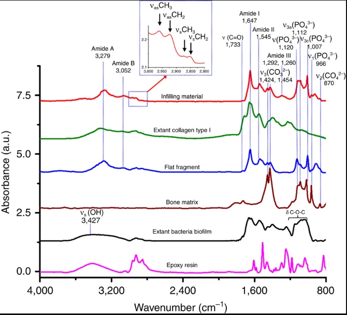
Baseline-corrected and normalized characteristic infrared band assignment for preserved collagen within the central vascular canals shown in red, and the peaks were assigned for methyl group (sCH3 and as CH3) and methylene (sCH2 and as CH2) in the spectral range of 3,000–2,800 cm−1 as shown in the blue inset, collagen type I from extant calf skin dispersed in 0.1% acetic acid solution in green, preserved protein remains in flat fragments found in and near the central canals of the fossil bone in blue, bone matrix in brown, extant bacteria biofilm in black and epoxy resin in pink. It is evident that the spectra of preserved collagen and extant collagen type I are closely matched. The extant bacterial biofilm showed significant differences from fossil or extant collagen in the range of 3,100–3,600 cm−1 region (sOH).
“Interestingly, there was no evidence of preservation of organic remains in the main mass of the bone, only in the small vascular canals that ran along the length of the rib, where hematite was also present” says Reisz.
“Our localized search, in areas of the bone that are likely to preserve remnants of the original soft tissues, is more likely to succeed than previously used methods. This approach has great future potential, because localized searches will yield important results even when the amount of organic remains is miniscule.”
Previous evidence of preserved collagen date back to the Late Cretaceous Period — more than 100 million years younger than this discovery — but those studies extracted the organic remains by dissolving away all other parts of the fossil, without a clear understanding of the precise origins of the collagen.
This research allowed the scientists to find the collagen in place without dissolving the rest of the fossil, and it has helped them understand how the organic remains were preserved. Reisz believes that future explorations for even older proteins will be possible if this technique is used.
@WFS,World Fossil Society,Riffin T Sajeev,Russel T Sajeev
 Posted in
  Tags:
Tags: 