

 September 24th, 2023
  Riffin
 @WFS,World Fossil Society, Athira, Riffin T Sajeev,Russel T Sajeev
A 455-million-year-old fossil fish provides a new perspective on how vertebrates evolved to protect their brains, a study has found.
In a paper published in Nature today (Wednesday 20th September), researchers from the University of Birmingham, Naturalis Biodiversity Centre in Leiden, Netherlands; and the Natural History Museum have pieced together the skull of Eriptychius americanus.
The research, funded by the Leverhulme Trust, suggests that the ancient jawless fish found in ancient deposits in Colorado, USA has a skull unlike that of any previously seen, and fills a gap currently spanning 100 million years in the evolutionary history of the vertebrate skull.
Using computed tomography, a form of x-ray technique, scientists recreated a detailed 3D representation of the skull of Eriptychius and is the first time that such a comprehensive recreation has been done on the specimen which was collected in the 1940s, originally described in the 1960s and is housed in the Field Museum of Natural History, Chicago.
This ancient fish had separated, independent cartilages encasing the brain, rather than the solid bone or cartilage structure of jawless and jawed fish that followed it.
While later specieshave a fully bound cage of cartilage that holds the brain, these results suggest that the early evolution of structures to separate the brain from other parts of the head may have begun with Eriptychius.
Dr Ivan Sansom, Senior Lecturer in Palaeobiology at the University of Birmingham and senior author of the paper said:
“These are tremendously exciting results that may reveal the early evolutionary history of how primitive vertebrates protected their brains. Eriptychius americanus appears to be the first evidence for a series of cartilages separating the brain from the rest of the head. This study emphasises the importance of museum collections and the application of new techniques in studying them.”
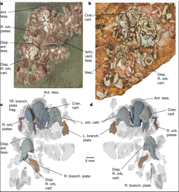
a,b, Photographs of part PF 1795a, which had the split face set in epoxy and was manually prepared (a), and its counterpart PF 1795b, which remains in rock matrix (b). Both are shown in an anatomically ventral view. c,d, Digital model of computed tomographic data of the combined part and counterpart with most of dermal skeleton rendered transparent: anatomical ventral view (corresponding to the visible area of the part in epoxy) (c) and anatomical dorsal view (buried in matrix in the counterpart) (d). Colour scheme for renders: blue-greys, cranial cartilages (matching the detailed scheme in Fig. 2); transparencies, the dermal skeleton; orange, branchial plates; red, orbital plates. Anterior to top in a–d. ant. tess., anterior tesserae; artic. vent. tess., articulated ventral tesserae; branch. plate, branchial plate; cran. cart., cranial cartilages; disp., displaced; frag., fragment; L., left; orb. cart., orbital cartilage; orb. plates, orbital plates; R., right; vasc., vasculature; ?, probable. Scale bar applies to all panels.
Dr Richard Dearden, Postdoctoral Research Fellow in Palaeobiology at Naturalis Biodiversity Center and lead author of the paper said:
“On the face of it Eriptychius is not the most beautiful of fossils. However, by using modern imaging techniques we were able to show that it preserves something unique: the oldest three-dimensionally preserved vertebrate head in the fossil record. This fills a major gap in our understanding of the evolution of the skull of all vertebrates, ultimately including humans.”
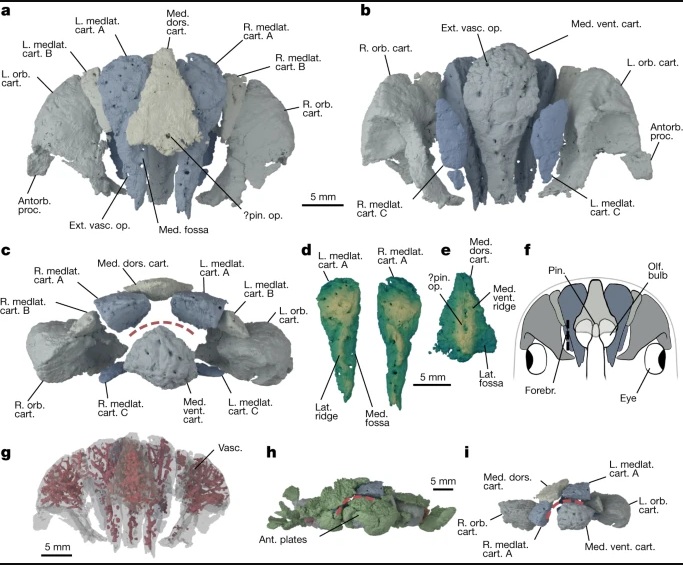
a–c, Cranial cartilages in estimated life position, with cartilages coloured in pairs in dorsal (a), ventral (b) and anterior (c) view. d,e, Mediolateral cartilages A in dorsal view (d) and median dorsal cartilage in ventral view (e) rendered with a vertical height map texture to emphasize the surface topology. f, Reconstruction of the forebrain relative to the cranial cartilages using a lamprey as a model9,52, shown in dorsal view. g, Cartilages in dorsal view, rendered transparent to show internal vasculature (red). h,i, Cartilages in preserved position in anterior view with dermal skeleton shown (h) and removed (i). Colours in a,b,c,f,h,i as in Fig. 1 with the following additions. Green, dermal skeleton. Red dashed line represents inferred position of mouth in c,h,i. In d and e lighter colours denote areas closer to the camera. Abbreviations as in Fig. 1 with the following additions: antorb. proc, antorbital process; ext. vasc. op., external vascular openings; forebr., forebrain; lat., lateral; medlat. cart, mediolateral cartilage; med. dors. cart, median dorsal cartilage; med., medial; med. vent. cart., median ventral cartilage; med. vent. ridge, median ventral ridge; olf. bulb, olfactory bulb; pin., pineal organ; pin. op., pineal opening; vent., ventral. Scale bar in a is shared by b,c; scale bar in d is shared by e.
- Dearden, R.P., Lanzetti, A., Giles, S. et al. The oldest three-dimensionally preserved vertebrate neurocranium. Nature, 2023 DOI: 10.1038/s41586-023-06538-y
 Posted in
  Tags:
Tags: 