WFS News: The oldest three-dimensionally preserved vertebrate neurocranium.

@WFS,World Fossil Society, Athira, Riffin T Sajeev,Russel T Sajeev

A 455-million-year-old fossil fish provides a new perspective on how vertebrates evolved to protect their brains, a study has found.

In a paper published in Nature today (Wednesday 20th September), researchers from the University of Birmingham, Naturalis Biodiversity Centre in Leiden, Netherlands; and the Natural History Museum have pieced together the skull of Eriptychius americanus.

The research, funded by the Leverhulme Trust, suggests that the ancient jawless fish found in ancient deposits in Colorado, USA has a skull unlike that of any previously seen, and fills a gap currently spanning 100 million years in the evolutionary history of the vertebrate skull.

Using computed tomography, a form of x-ray technique, scientists recreated a detailed 3D representation of the skull of Eriptychius and is the first time that such a comprehensive recreation has been done on the specimen which was collected in the 1940s, originally described in the 1960s and is housed in the Field Museum of Natural History, Chicago.

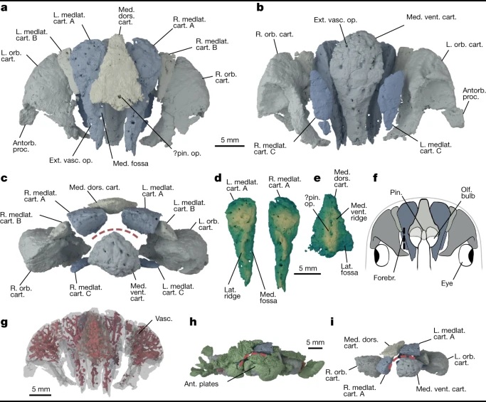
This ancient fish had separated, independent cartilages encasing the brain, rather than the solid bone or cartilage structure of jawless and jawed fish that followed it.

While later specieshave a fully bound cage of cartilage that holds the brain, these results suggest that the early evolution of structures to separate the brain from other parts of the head may have begun with Eriptychius.

Dr Ivan Sansom, Senior Lecturer in Palaeobiology at the University of Birmingham and senior author of the paper said:

“These are tremendously exciting results that may reveal the early evolutionary history of how primitive vertebrates protected their brains. Eriptychius americanus appears to be the first evidence for a series of cartilages separating the brain from the rest of the head. This study emphasises the importance of museum collections and the application of new techniques in studying them.”

a,b, Photographs of part PF 1795a, which had the split face set in epoxy and was manually prepared (a), and its counterpart PF 1795b, which remains in rock matrix (b). Both are shown in an anatomically ventral view. c,d, Digital model of computed tomographic data of the combined part and counterpart with most of dermal skeleton rendered transparent: anatomical ventral view (corresponding to the visible area of the part in epoxy) (c) and anatomical dorsal view (buried in matrix in the counterpart) (d). Colour scheme for renders: blue-greys, cranial cartilages (matching the detailed scheme in Fig. 2); transparencies, the dermal skeleton; orange, branchial plates; red, orbital plates. Anterior to top in a–d. ant. tess., anterior tesserae; artic. vent. tess., articulated ventral tesserae; branch. plate, branchial plate; cran. cart., cranial cartilages; disp., displaced; frag., fragment; L., left; orb. cart., orbital cartilage; orb. plates, orbital plates; R., right; vasc., vasculature; ?, probable. Scale bar applies to all panels.

a,b, Photographs of part PF 1795a, which had the split face set in epoxy and was manually prepared (a), and its counterpart PF 1795b, which remains in rock matrix (b). Both are shown in an anatomically ventral view. c,d, Digital model of computed tomographic data of the combined part and counterpart with most of dermal skeleton rendered transparent: anatomical ventral view (corresponding to the visible area of the part in epoxy) (c) and anatomical dorsal view (buried in matrix in the counterpart) (d). Colour scheme for renders: blue-greys, cranial cartilages (matching the detailed scheme in Fig. 2); transparencies, the dermal skeleton; orange, branchial plates; red, orbital plates. Anterior to top in a–d. ant. tess., anterior tesserae; artic. vent. tess., articulated ventral tesserae; branch. plate, branchial plate; cran. cart., cranial cartilages; disp., displaced; frag., fragment; L., left; orb. cart., orbital cartilage; orb. plates, orbital plates; R., right; vasc., vasculature; ?, probable. Scale bar applies to all panels.

Dr Richard Dearden, Postdoctoral Research Fellow in Palaeobiology at Naturalis Biodiversity Center and lead author of the paper said:

“On the face of it Eriptychius is not the most beautiful of fossils. However, by using modern imaging techniques we were able to show that it preserves something unique: the oldest three-dimensionally preserved vertebrate head in the fossil record. This fills a major gap in our understanding of the evolution of the skull of all vertebrates, ultimately including humans.”

a–c, Cranial cartilages in estimated life position, with cartilages coloured in pairs in dorsal (a), ventral (b) and anterior (c) view. d,e, Mediolateral cartilages A in dorsal view (d) and median dorsal cartilage in ventral view (e) rendered with a vertical height map texture to emphasize the surface topology. f, Reconstruction of the forebrain relative to the cranial cartilages using a lamprey as a model9,52, shown in dorsal view. g, Cartilages in dorsal view, rendered transparent to show internal vasculature (red). h,i, Cartilages in preserved position in anterior view with dermal skeleton shown (h) and removed (i). Colours in a,b,c,f,h,i as in Fig. 1 with the following additions. Green, dermal skeleton. Red dashed line represents inferred position of mouth in c,h,i. In d and e lighter colours denote areas closer to the camera. Abbreviations as in Fig. 1 with the following additions: antorb. proc, antorbital process; ext. vasc. op., external vascular openings; forebr., forebrain; lat., lateral; medlat. cart, mediolateral cartilage; med. dors. cart, median dorsal cartilage; med., medial; med. vent. cart., median ventral cartilage; med. vent. ridge, median ventral ridge; olf. bulb, olfactory bulb; pin., pineal organ; pin. op., pineal opening; vent., ventral. Scale bar in a is shared by b,c; scale bar in d is shared by e.

a–c, Cranial cartilages in estimated life position, with cartilages coloured in pairs in dorsal (a), ventral (b) and anterior (c) view. d,e, Mediolateral cartilages A in dorsal view (d) and median dorsal cartilage in ventral view (e) rendered with a vertical height map texture to emphasize the surface topology. f, Reconstruction of the forebrain relative to the cranial cartilages using a lamprey as a model9,52, shown in dorsal view. g, Cartilages in dorsal view, rendered transparent to show internal vasculature (red). h,i, Cartilages in preserved position in anterior view with dermal skeleton shown (h) and removed (i). Colours in a,b,c,f,h,i as in Fig. 1 with the following additions. Green, dermal skeleton. Red dashed line represents inferred position of mouth in c,h,i. In d and e lighter colours denote areas closer to the camera. Abbreviations as in Fig. 1 with the following additions: antorb. proc, antorbital process; ext. vasc. op., external vascular openings; forebr., forebrain; lat., lateral; medlat. cart, mediolateral cartilage; med. dors. cart, median dorsal cartilage; med., medial; med. vent. cart., median ventral cartilage; med. vent. ridge, median ventral ridge; olf. bulb, olfactory bulb; pin., pineal organ; pin. op., pineal opening; vent., ventral. Scale bar in a is shared by b,c; scale bar in d is shared by e.

  1. Dearden, R.P., Lanzetti, A., Giles, S. et al. The oldest three-dimensionally preserved vertebrate neurocraniumNature, 2023 DOI: 10.1038/s41586-023-06538-y
University of Birmingham. “Prehistoric fish fills 100 million year gap in evolution of the skull.” ScienceDaily. ScienceDaily, 20 September 2023. <www.sciencedaily.com/releases/2023/09/230920110303.htm>.
@WFS,World Fossil Society, Athira, Riffin T Sajeev,Russel T Sajeev,

WFS News: Rhabdodontidae dinosaurs of Late Cretaceous Europe

@WFS,World Fossil Society, Athira, Riffin T Sajeev,Russel T Sajeev

A new study published in Fossil Record brings together intriguing details about the little-known Rhabdodontidae dinosaurs of Late Cretaceous Europe. These gregarious herbivores, characterized by robust builds and beaks specialized for tough vegetation, inhabited the European archipelago. Despite being widespread and abundant, they vanished in Western Europe due to environmental changes around 69 million years ago, while surviving longer in Eastern Europe. Their fossil record offers valuable insights into their evolution and lifestyle, although its limited nature still challenges comprehensive understanding.

When you think of dinosaurs, you might automatically imagine iconic dinosaurs as Tyrannosaurus and Triceratops. But at the same time when these were stomping on the ancient coastal plains of North America, some of their very distant cousins were reigning over Europe’s lands.

During the Late Cretaceous (between 100 and 66 million years ago), Europe was an extensive archipelago with numerous small and large islands situated in a shallow tropical sea, the so-called Late Cretaceous European Archipelago. The dinosaur groups that lived on these islands were very different from those of other continents, often being much smaller than their mainland relatives. These European dinosaurs include small and medium-sized carnivorous theropods, armoured ankylosaurs, long-necked sauropods, duck-billed hadrosaurs, and rhabdodontids.

Arguably one of the most important of these European dinosaur groups is the family Rhabdodontidae, which groups together the most common medium-sized herbivores of the Late Cretaceous European Archipelago. A joint research team from the Universities of Tübingen (Germany), Budapest (Hungary) and Bucharest (Romania) recently reviewed what we know about these peculiar dinosaurs in a new paper published in the journal Fossil Record.

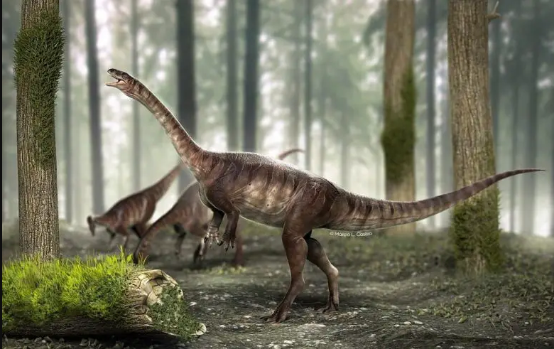
Generally, rhabdodontid dinosaurs were small to medium-sized animals with an overall body length of approximately 2-6 m. “They were probably habitually bipedal herbivores, characterised by a rather stocky build, with strong hind limbs, short forelimbs, a long tail, and a comparatively large, triangular skull that tapers anteriorly and ends in a narrow snout,” explains Felix Augustin, lead author of the study in Fossil Record.

“They had a relatively robust skull with strong jaws, large teeth and a pointy beak that was covered in keratin, demonstrating that these dinosaurs were well-adapted to eating tough plants.”

In some instances, fossil remains of several individuals of different ages have been found together, indicating that they were gregarious.

Although they died out well before the mass extinction in Western Europe (about 69 million years ago), potentially due to environmental changes that affected the plants they fed on, they survived much longer in Eastern Europe and were among the last non-avian dinosaurs still present before the end of the Cretaceous (66 million years ago).

Interestingly, fossils of rhabdodontids have only been found in Europe and only in rocks ranging in age from 86-66 million years ago, so they were endemic to the Late Cretaceous European Archipelago.

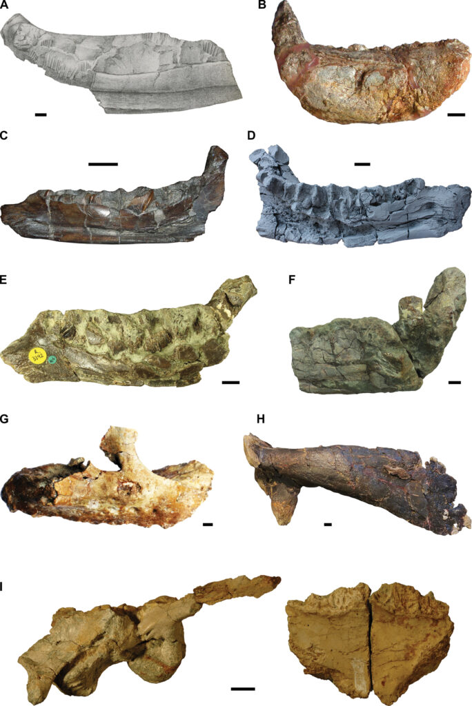
Type specimens of the nine rhabdodontid species described so far. A. The original drawing of the lectotype of Rhabdodon priscus, MPLM 30, a partial left dentary. The specimen has since deteriorated (Pincemaille-Quillevere 2002). Modified after Matheron (1869). B. Holotype of Rhabdodon septimanicus, MDE D-30, an incomplete right dentary. Photo kindly provided by Eric Buffetaut. C. Lectotype of Mochlodon suessi, PIUW 2349/2, a right dentary. D. Holotype of Mochlodon vorosi, MTM V 2010.105.1, a left dentary. E. Holotype of Zalmoxes robustus, NHMUK R.3392, a right dentary. Photo kindly provided by János Magyar. F. Holotype right dentary of Zalmoxes shqiperorum, NHMUK R.4900. Note that the holotype of Z. shqiperorum also comprises several postcranial elements that presumably belong to the same individual as the dentary. Photo kindly provided by János Magyar. G. Holotype of Matheronodon provincialis, MMS/VBN-02-102, a right maxilla. Modified after Godefroit et al. (2017). H. Holotype of Pareisactus evrostos, MCD 5371, a left scapula. Modified after Párraga and Prieto-Márquez (2019). I. Holotype of Transylvanosaurus platycephalus, LPB (FGGUB) R.2070, a partial skull comprising the articulated basicranium and both frontals. Scale bars: 1 cm.

Type specimens of the nine rhabdodontid species described so far. A. The original drawing of the lectotype of Rhabdodon priscus, MPLM 30, a partial left dentary. The specimen has since deteriorated (Pincemaille-Quillevere 2002). Modified after Matheron (1869). B. Holotype of Rhabdodon septimanicus, MDE D-30, an incomplete right dentary. Photo kindly provided by Eric Buffetaut. C. Lectotype of Mochlodon suessi, PIUW 2349/2, a right dentary. D. Holotype of Mochlodon vorosi, MTM V 2010.105.1, a left dentary. E. Holotype of Zalmoxes robustus, NHMUK R.3392, a right dentary. Photo kindly provided by János Magyar. F. Holotype right dentary of Zalmoxes shqiperorum, NHMUK R.4900. Note that the holotype of Z. shqiperorum also comprises several postcranial elements that presumably belong to the same individual as the dentary. Photo kindly provided by János Magyar. G. Holotype of Matheronodon provincialis, MMS/VBN-02-102, a right maxilla. Modified after Godefroit et al. (2017). H. Holotype of Pareisactus evrostos, MCD 5371, a left scapula. Modified after Párraga and Prieto-Márquez (2019). I. Holotype of Transylvanosaurus platycephalus, LPB (FGGUB) R.2070, a partial skull comprising the articulated basicranium and both frontals. Scale bars: 1 cm.

The group currently comprises nine different species from five European countries (France, Spain, Austria, Hungary, and Romania).

“The first rhabdodontid species was scientifically named more than 150 years ago and the last one as recently as November 2022, so, although the group looks back to a long research history, we still have much to learn about it,” says Felix Augustin.

“Generally, our portraying of the world of dinosaurs is heavily biased towards the well-known North-American and Asian dinosaur faunas,” he adds.

Dinosaur fossils from the Late Cretaceous are much rarer in Europe than in North America or Asia, and thus far no complete skeleton of a rhabdodontid has been described. Even though they were so abundant and common in the Upper Cretaceous of Europe, several key aspects about them remain poorly known, including their detailed body proportions, their posture and locomotion, as well as their feeding behaviour.

“In the past decades, a wealth of new, and often well-preserved, rhabdodontid fossils has been discovered throughout Europe, the majority of which still remains to be studied,” says Felix Augustin. “A joint research project is currently underway to study the available fossil material in order to gain new insights into the evolution and lifestyle of these fascinating yet still poorly known dinosaurs.”

  1. Felix J. Augustin, Attila Ősi, Zoltán Csiki-Sava. The Rhabdodontidae (Dinosauria, Ornithischia), an enigmatic dinosaur group endemic to the Late Cretaceous European ArchipelagoFossil Record, 2023; 26 (2): 171 DOI: 10.3897/fr.26.108967
Pensoft Publishers. “Europe’s very own dinosaurs — the enigmatic Late Cretaceous rhabdodontids.” ScienceDaily. ScienceDaily, 1 September 2023. <www.sciencedaily.com/releases/2023/08/230830131713.htm>.
@WFS,World Fossil Society, Athira, Riffin T Sajeev,Russel T Sajeev

WFS News: Sulfur minerals that make fossils are especially well-suited to radiography

@WFS,World Fossil Society, Athira, Riffin T Sajeev,Russel T Sajeev

New research reveals that sulfur minerals that make fossils in the Norwegian archipelago are especially well-suited to radiography.

An overlook of the Muen plateau as seen from the Muen mountain on Edgeøya, Svalbard. Marine reptiles are spread out across the plateau. Author VSE in red jacket for scale. Credit: Sofie Bernhardsen, CC-BY 4.0

An overlook of the Muen plateau as seen from the Muen mountain on Edgeøya, Svalbard. Marine reptiles are spread out across the plateau. Author VSE in red jacket for scale. Credit: Sofie Bernhardsen, CC-BY 4.0

X-ray analysis has led to the categorization of a previously-unidentified marine reptile fossil discovered in Edgeøya, Svalbard. The research was recently published in the journal PLOS ONE.  The study, conducted by Victoria S. Engelschiøn of the University of Oslo and her team, suggests that this method could reveal fresh insights about ancient life in the future.

The effectiveness of X-ray techniques in investigating well-conserved fossil remains is often dependent on the condition of preservation, which can vary greatly across different sites. Through this study, Engelschiøn and her team showed that fossils from the Middle Triassic Botneheia Formation in Svalbard, Norway, are particularly suitable for radiographic imaging.

The focus of this study is a fossil marine reptile whose remains are compressed and encased in shale. It lived around 240 million years ago, when Svalbard was covered by an ocean. After it died, it sank to the seafloor and was buried in the mud, then became extremely flattened over time. Originally excavated in 2008, the identity of this fossil has since been debated. X-ray imaging of the specimen revealed new details, including features of the skull and teeth that allowed researchers to conclude that this reptile most likely belongs to the ichthyosaur species Phalarodon atavus.

Photograph of specimen PMO 219.250. Credit: Engelschiøn et al. PLOS ONE, CC-BY 4.0

Photograph of specimen PMO 219.250. Credit: Engelschiøn et al. PLOS ONE, CC-BY 4.0

The authors also examined the mineralogy of fossils from this formation, identifying multiple forms of sulfate minerals, notably including sulfate baryte, which gives the fossils very high X-ray contrast, allowing for the high quality of radiographic imaging. The formation of these minerals is little understood but could be linked to conditions created by ancient volcanic activity. Thus, this study not only demonstrates the utility of X-ray techniques for studying these fossils, but also identifies conditions that can form fossils well-suited for these techniques, in Svalbard and potentially elsewhere.

The authors add: “The rocks from Svalbard are full of flattened marine reptiles. Our discovery of the exceptional X-Ray contrast means that we can learn much more about these ancient predators than we previously thought.”

Reference: “Exceptional X-Ray contrast: Radiography imaging of a Middle Triassic mixosaurid from Svalbard” by Victoria S. Engelschiøn, Aubrey J. Roberts, Ruben With and Øyvind Hammer, 31 May 2023, PLOS ONE.
DOI: 10.1371/journal.pone.0285939

@WFS,World Fossil Society, Athira, Riffin T Sajeev,Russel T Sajeev

WFS News: New insights into the sea spider fauna (Arthropoda, Pycnogonida) of La Voulte‐sur‐Rhône, France (Jurassic, Callovian)

@WFS,World Fossil Society, Athira, Riffin T Sajeev,Russel T Sajeev

An extremely rare collection of 160-million-year-old sea spider fossils from Southern France are closely related to living species, unlike older fossils of their kind.

These fossils are very important to understand the evolution of sea spiders. They show that the diversity of sea spiders that still exist today had already started to form by the Jurassic.

Lead author Dr Romain Sabroux from the University of Bristol’s School of Earth Sciences, said: “Sea spiders (Pycnogonida), are a group of marine animals that is overall very poorly studied.

“However, they are very interesting to understand the evolution of arthropods [the group that includes insects, arachnids, crustaceans, centipedes and millipedes] as they appeared relatively early in the arthropod tree of life. That’s why we are interested in their evolution.

“Sea spider fossils are very rare, but we know a few of them from different periods. One of the most remarkable fauna, by its diversity and its abundance, is the one of La Voulte-sur-Rhône that dates back to the Jurassic, some 160 million years ago.”

Unlike older sea spider fossils, the La Voulte pycnogonids are morphologically similar (but not identical) to living species, and previous studies suggested they could be closely related to living sea spider families. But these hypotheses were restricted by the limitation of their observation means. As it was impossible to access what was hidden in the rock fossils, Dr Sabroux and his team travelled to Paris and set out to investigate this question with cutting-edge approaches.

RTI of Palaeopycnogonides gracilis, MNHN.F.A88075, body region (preserved ventrally, with imprint of dorsal region anteriorly). A, default view. B, specular enhancement (circle on bottom right indicates light orientation, see Table S2 for details). C, ‘normals visualization’. D, interpretative drawing; plain black lines correspond to the outline of the fossil; dashed black lines correspond to the specimen imprints; dotted black lines correspond to the putative ovigers; grey lines correspond to the main breaks in the fossil. Abbreviations: an, anus; gp, gonopore. Scale bars represent 5 mm.

RTI of Palaeopycnogonides gracilis, MNHN.F.A88075, body region (preserved ventrally, with imprint of dorsal region anteriorly). A, default view. B, specular enhancement (circle on bottom right indicates light orientation, see Table S2 for details). C, ‘normals visualization’. D, interpretative drawing; plain black lines correspond to the outline of the fossil; dashed black lines correspond to the specimen imprints; dotted black lines correspond to the putative ovigers; grey lines correspond to the main breaks in the fossil. Abbreviations: an, anus; gp, gonopore. Scale bars represent 5 mm.

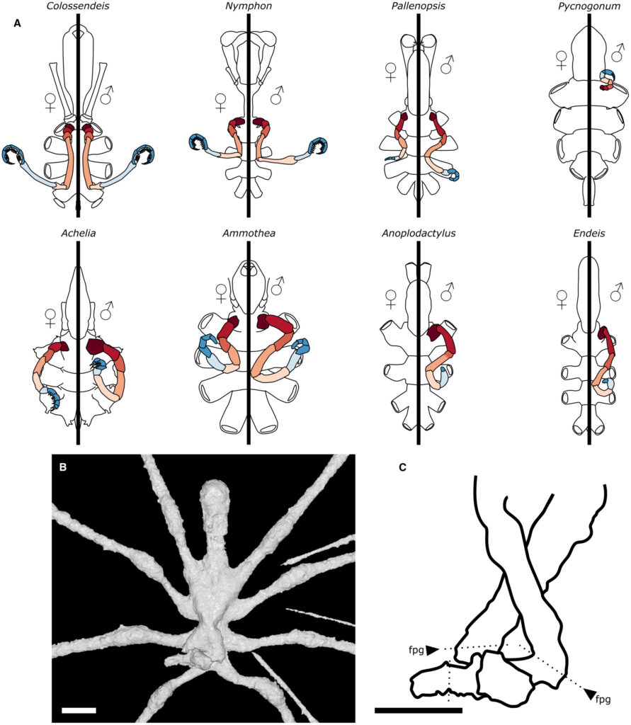
Dr Sabroux explained: “We used two methods to reinvestigate the morphology of the fossils: X-ray microtomography, to ‘look inside’ the rock, find morphological features hidden inside and reconstruct a 3D model of the fossilised specimen; and Reflectance Transformation Imaging, a picture technic that relies on varied orientation of the light around the fossil to enhance the visibility of inconspicuous features on their surface.

“From these new insights, we drew new morphological information to compare them with extant species,” explained Dr Sabroux.

This confirmed that these fossils are close relatives to surviving pycnogonids. Two of these fossils belong to two living pycnogonid families: Colossopantopodus boissinensis was a Colossendeidae while another, Palaeoendeis elmii was an Endeidae. The third species, Palaeopycnogonides gracilis, seems to belong to a family that has disappeared today.

“Today, by calculating the difference between the DNA sequences of a sample of species, and using DNA evolution models, we are able to estimate the timing of the evolution that bind these species together,” added Dr Sabroux.

“This is what we call a molecular clock analysis. But quite like a real clock, it needs to be calibrated. Basically, we need to tell the clock: ‘we know that at that time, that group was already there.’ Thanks to our work, we now know that Colossendeidae, and Endeidae were already ‘there’ by the Jurassic.”

Now, the team can use these minimal ages as calibrations for the molecular clock, and investigate the timing of Pycnogonida evolution. This can help them understand, for example, how their diversity was impacted by the different biodiversity crises that distributes over the Earth history.

They also plan to investigate other pycnogonid fossil faunae such as the fauna of Hunsrück Slate, in Germany, which dates from the Devonian, some 400 million years ago.

With the same approach, they will aim to redescribe these species and understand their affinities with extant species; and finally, to replace in the tree of life of Pycnogonida all the pycnogonid fossils from all periods.

Comparison of the ovigers of Palaeoendeis elmii with some extant pantopods. A, ovigers as found in the two sexes in eight examples of modern sea spiders, using the same colour code as in Figure 1. B, close-up of the ventral view of the three-dimensional reconstruction of P. elmii, holotype MNHN.F.A49277. C, interpretative drawing of the ovigers of P. elmii; plain black lines correspond to the outline of the fossil; dotted lines correspond to the putative position of articulations. Abbreviation: fpg, femoro-patellar geniculation. Scale bars represent 2 mm.

Comparison of the ovigers of Palaeoendeis elmii with some extant pantopods. A, ovigers as found in the two sexes in eight examples of modern sea spiders, using the same colour code as in Figure 1. B, close-up of the ventral view of the three-dimensional reconstruction of P. elmii, holotype MNHN.F.A49277. C, interpretative drawing of the ovigers of P. elmii; plain black lines correspond to the outline of the fossil; dotted lines correspond to the putative position of articulations. Abbreviation: fpg, femoro-patellar geniculation. Scale bars represent 2 mm.

Dr Sabroux added: “These fossils give us an insight of sea spiders living 160 million years ago.

“This is very exciting when you have been working on the living pycnogonids for years.

“It is fascinating how these pycnogonids look both very familiar, and very exotic. Familiar, because you can definitely recognize some of the families that still exist today, and exotic because of small differences like the size of the legs, the length of the body, and some other morphological characteristics that you do not find in modern species.

“Now we look forward to the next fossil discoveries — from the Jurassic and other geological periods — so that we can complete the picture!”

Journal Reference:

  1. Romain Sabroux, Gregory D. Edgecombe, Davide Pisani, Russell J. Garwood. New insights into the sea spider fauna (Arthropoda, Pycnogonida) of La Voulte‐sur‐Rhône, France (Jurassic, Callovian)Papers in Palaeontology, 2023; 9 (4) DOI: 10.1002/spp2.1515
University of Bristol. “The modern sea spider had started to diversify by the Jurassic, study finds.” ScienceDaily. ScienceDaily, 17 August 2023. <www.sciencedaily.com/releases/2023/08/230817164019.htm>.
@WFS,World Fossil Society, Athira, Riffin T Sajeev,Russel T Sajeev


WFS News:Burgessomedusa phasmiformis;Oldest known species of swimming jellyfish

@WFS,World Fossil Society, Athira, Riffin T Sajeev,Russel T Sajeev

Life reconstruction showing a cluster of Burgessomedusa phasmiformis gen. et sp. nov. swimming above the benthos. This reconstruction is based on the Raymond Quarry Burgess Shale community with clusters of Vauxia sponges represented in the foreground. Artwork by C. McCall.

Life reconstruction showing a cluster of Burgessomedusa phasmiformis gen. et sp. nov. swimming above the benthos. This reconstruction is based on the Raymond Quarry Burgess Shale community with clusters of Vauxia sponges represented in the foreground. Artwork by C. McCall.

Royal Ontario Museum (ROM) announces the oldest swimming jellyfish in the fossil record with the newly named Burgessomedusa phasmiformis. These findings are announced in the journal Proceedings of the Royal Society B.

Jellyfish belong to medusozoans, or animals producing medusae, and include today’s box jellies, hydroids, stalked jellyfish and true jellyfish. Medusozoans are part of one of the oldest groups of animals to have existed, called Cnidaria, a group which also includes corals and sea anemones. Burgessomedusa unambiguously shows that large, swimming jellyfish with a typical saucer or bell-shaped body had already evolved more than 500 million years ago.

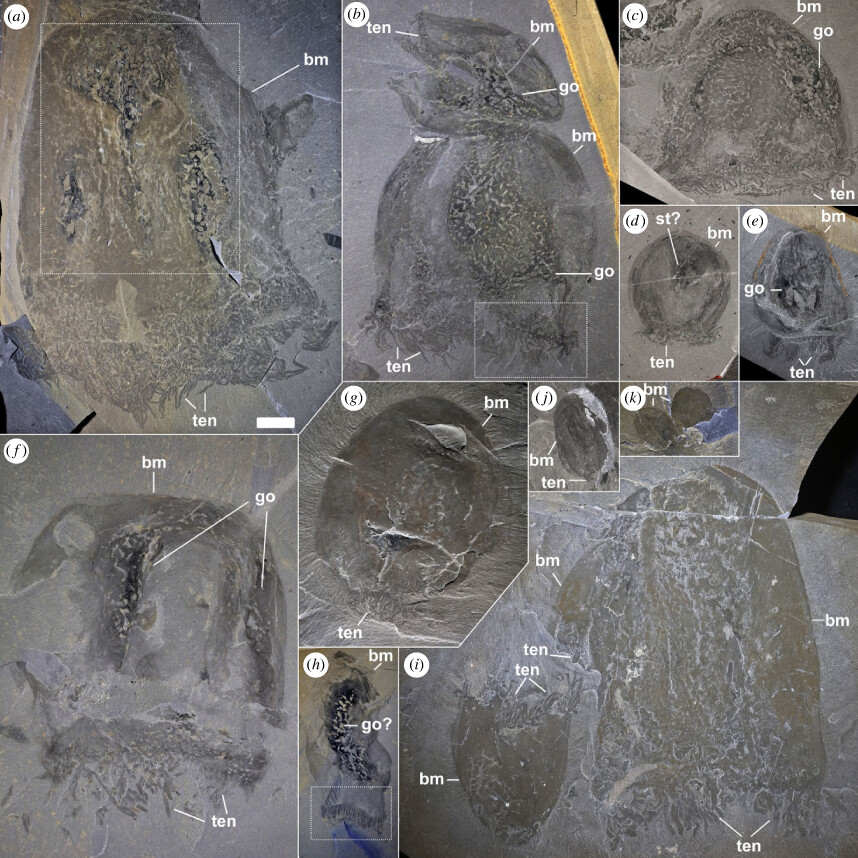
Size variations and general morpho-anatomical details of Burgessomedusa phasmiformis gen. et sp. nov. (a) Holotype ROMIP65781.1 (close-up in figure 2a). (b) ROMIP65782.2–3, with putative gonads (close-up in figure 2b). (c), ROMIP65783.1, with putative gonads. (d) ROMIP65784, with putative stomach cavity. e,f, specimens with putative gonads ROMIP65785 (e), ROMIP65786 (f). (g) ROMIP65787, with a contracted umbrella. (h) ROMIP65788, with putative gonads (close-up in figure 2e). (i) ROMIP65114.1–3. (j) ROMIP65789. (k) ROMIP65790.1–2. Abbreviations: bm, bell margin; go, gonads; man, manubrium; st, stomach cavity; ten, tentacles. Scale = 2 cm.

Size variations and general morpho-anatomical details of Burgessomedusa phasmiformis gen. et sp. nov. (a) Holotype ROMIP65781.1 (close-up in figure 2a). (b) ROMIP65782.2–3, with putative gonads (close-up in figure 2b). (c), ROMIP65783.1, with putative gonads. (d) ROMIP65784, with putative stomach cavity. e,f, specimens with putative gonads ROMIP65785 (e), ROMIP65786 (f). (g) ROMIP65787, with a contracted umbrella. (h) ROMIP65788, with putative gonads (close-up in figure 2e). (i) ROMIP65114.1–3. (j) ROMIP65789. (k) ROMIP65790.1–2. Abbreviations: bm, bell margin; go, gonads; man, manubrium; st, stomach cavity; ten, tentacles. Scale = 2 cm.

Burgessomedusa fossils are exceptionally well preserved at the Burgess Shale considering jellyfish are roughly 95% composed of water. ROM holds close to two hundred specimens from which remarkable details of internal anatomy and tentacles can be observed, with some specimens reaching more than 20 centimetres in length. These details enable classifying Burgessomedusa as amedusozoan. By comparison with modern jellyfish, Burgessomedusa would also have been capable of free-swimming and the presence of tentacles would have enabled capturing sizeable prey.

“Although jellyfish and their relatives are thought to be one of the earliest animal groups to have evolved, they have been remarkably hard to pin down in the Cambrian fossil record. This discovery leaves no doubt they were swimming about at that time,” said co-author Joe Moysiuk, a Ph.D. candidate in Ecology & Evolutionary Biology at the University of Toronto, who is based at ROM.

This study, identifying Burgessomedusa, is based on fossil specimens discovered at the Burgess Shale and mostly found in the late 1980s and 1990s under former ROM Curator of Invertebrate Palaeontology Desmond Collins. They show that the Cambrian food chain was far more complex than previously thought, and that predation was not limited to large swimming arthropods like Anomalocaris (see field image showing Burgessomedusa and Anomalocaris preserved on the same rock surface).

“Finding such incredibly delicate animals preserved in rock layers on top of these mountains is such a wonderous discovery. Burgessomedusa adds to the complexity of Cambrian foodwebs, and like Anomalocaris which lived in the same environment, these jellyfish were efficient swimming predators,” said co-author, Dr. Jean-Bernard Caron, ROM’s Richard Ivey Curator of Invertebrate Palaeontology. “This adds yet another remarkable lineage of animals that the Burgess Shale has preserved chronicling the evolution of life on Earth.”

Cnidarians have complex life cycles with one or two body forms, a vase-shaped body, called a polyp, and in medusozoans, a bell or saucer-shaped body, called a medusa or jellyfish, which can be free-swimming or not. While fossilized polyps are known in ca. 560-million-year-old rocks, the origin of the free-swimming medusa or jellyfish is not well understood. Fossils of any type of jellyfish are extremely rare. As a consequence, their evolutionary history is based on microscopic fossilized larval stages and the results of molecular studies from living species (modelling of divergence times of DNA sequences). Though some fossils of comb-jellies have also been found at the Burgess Shale and in other Cambrian deposits, and may superficially resemble medusozoan jellyfish from the phylum Cnidaria, comb-jellies are actually from a quite separate phylum of animals called Ctenophora. Previous reports of Cambrian swimming jellyfish are reinterpreted as ctenophores.

The Burgess Shale fossil sites are located within Yoho and Kootenay National Parks and are managed by Parks Canada. Parks Canada is proud to work with leading scientific researchers to expand knowledge and understanding of this key period of Earth history and to share these sites with the world through award-winning guided hikes. The Burgess Shale was designated a UNESCO World Heritage Site in 1980 due to its outstanding universal value and is now part of the larger Canadian Rocky Mountain Parks World Heritage Site.

  1. Justin Moon, Jean-Bernard Caron, Joseph Moysiuk. A macroscopic free-swimming medusa from the middle Cambrian Burgess ShaleProceedings of the Royal Society B: Biological Sciences, 2023; 290 (2004) DOI: 10.1098/rspb.2022.2490
Royal Ontario Museum. “Oldest known species of swimming jellyfish identified: 505-million-year-old swimming jellyfish from the Burgess Shale highlights diversity in Cambrian ecosystem.” ScienceDaily. ScienceDaily, 1 August 2023. <www.sciencedaily.com/releases/2023/08/230801200756.htm>.
@WFS,World Fossil Society, Athira, Riffin T Sajeev,Russel T Sajeev

WFS News: Newly discovered dinosaur, ‘Iani,’ was face of a changing planet

@WFS,World Fossil Society, Athira, Riffin T Sajeev,Russel T Sajeev

A newly discovered plant-eating dinosaur may have been a species’ “last gasp” during a period when Earth’s warming climate forced massive changes to global dinosaur populations.

The specimen, named Iani smithi after Janus, the two-faced Roman god of change, was an early ornithopod, a group of dinosaurs that ultimately gave rise to the more commonly known duckbill dinosaurs such as Parasaurolophus and Edmontosaurus. Researchers recovered most of the juvenile dinosaur’s skeleton—including skull, vertebrae and limbs—from Utah’s Cedar Mountain Formation. The research is published in PLoS ONE.

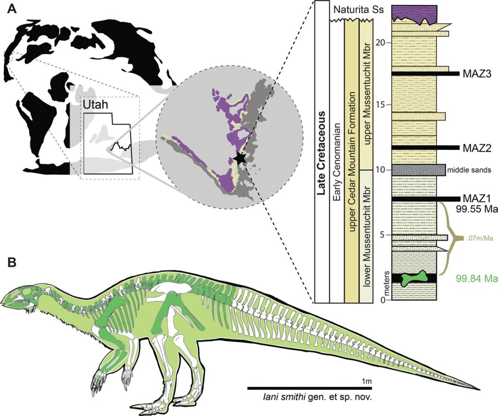
Location of holotype locality for Iani smithi. (A) Global map showing location of Mussentuchit Member outcrop in central Utah, western North America, and a stratigraphic section at the quarry with dated ash horizons; and (B) graphical representation of preserved skeletal elements of the holotype specimen. Preserved elements are colored on the left facing skeletal whether they derive from the right or left side of the body. Exact positions of chevrons and ribs unknown due to poor preservation. Credit: Zanno et al., 2023, PLOS ONE, CC-BY 4.0 (creativecommons.org/licenses/by/4.0/)

Location of holotype locality for Iani smithi. (A) Global map showing location of Mussentuchit Member outcrop in central Utah, western North America, and a stratigraphic section at the quarry with dated ash horizons; and (B) graphical representation of preserved skeletal elements of the holotype specimen. Preserved elements are colored on the left facing skeletal whether they derive from the right or left side of the body. Exact positions of chevrons and ribs unknown due to poor preservation. Credit: Zanno et al., 2023, PLOS ONE, CC-BY 4.0 (creativecommons.org/licenses/by/4.0/)

Iani smithi lived in what is now Utah during the mid-Cretaceous, approximately 99 million years ago. The dinosaur’s most striking feature is its powerful jaw, with teeth designed for chewing through tough plant material.

The mid-Cretaceous was a time of big changes, which had big effects on dinosaur populations. Increased  during this time caused the Earth to warm and sea levels to rise, corralling dinosaurs on smaller and smaller landmasses. It was so warm that rainforests thrived at the poles. Flowering plant life took over  and supplanted normal food sources for herbivores.

In North America, giant plant-eating sauropods—once titans of the landscape—were disappearing, along with their allosaurian predators. At the same time, smaller plant eaters, like early duckbills and horned dinosaurs, and feathered theropods like tyrannosaurs and enormous oviraptorosaurs, were arriving from Asia.

Enter Iani smithi, unique not only because it’s newly discovered, but also because of its rarity in the North American fossil record and its position in dinosaur history.

“Finding Iani was a streak of luck. We knew something like it lived in this ecosystem because isolated teeth had been collected here and there, but we weren’t expecting to stumble upon such a beautiful skeleton, especially from this time in Earth’s history. Having a nearly complete skull was invaluable for piecing the story together,” says Lindsay Zanno, associate research professor at North Carolina State University, head of paleontology at the North Carolina Museum of Natural Sciences, and corresponding author of the work.

Zanno and her team used the well-preserved skeleton to analyze the evolutionary relationships of Iani and were surprised—and a bit skeptical—of the results.

“We recovered Iani as an early rhabdodontomorph, a lineage of ornithopods known almost exclusively from Europe,” Zanno says. “Recently, paleontologists proposed that another North American dinosaur, Tenontosaurus—which was as common as cattle in the Early Cretaceous—belongs to this group, as well as some Australian critters. If Iani holds up as a rhabdodontomorph, it raises a lot of cool questions.”

Key among these is, could Iani be a last gasp, a witness to the end of a once successful lineage? Zanno thinks that studying this fossil in the context of environmental and biodiversity changes during the mid-Cretaceous will give us more insight into the history of our planet.

Iani smithi is named for Janus, the two-faced god who symbolized transitions—an apt name, given its position in history.

“Iani may be the last surviving member of a lineage of dinosaurs that once thrived here in North America but were eventually supplanted by duckbill dinosaurs,” Zanno says. “Iani was alive during this transition—so this dinosaur really does symbolize a changing planet.

“This dinosaur stood on the precipice,” she says, “able to look back at the way North American ecosystems were in the past, but close enough to see the future coming like a bullet train. I think we can all relate to that.”

Publication: An early-diverging iguanodontian (Dinosauria: Rhabdodontomorpha) from the Late Cretaceous of North America, PLoS ONE (2023). DOI: 10.1371/journal.pone.0286042

Source: Article by  in Phys,org

@WFS,World Fossil Society, Athira, Riffin T Sajeev,Russel T Sajeev

WFS News: Petrodactyle wellnhoferi gen. et sp. nov.: A new and large ctenochasmatid pterosaur from the Late Jurassic of Germany

@WFS,World Fossil Society, Athira, Riffin T Sajeev,Russel T Sajeev

A new 145-million-year-old pterosaur (extinct flying reptiles that lived alongside the dinosaurs) was named today by a team of British, American and German researchers. The animal was nicknamed ‘Elvis’ when the fossil was first unearthed in Bavaria, Germany because of the giant pompadour-like bony crest on its skull.

Now the animal has been given a formal scientific name of Petrodactyle wellnhoferi. The name translates as ‘Wellnhofer’s stone-finger’ honouring legendary German palaeontologist Peter Wellnhofer who spent his career working on German pterosaurs. Petrodactyle is a member of a group of pterosaurs called the ctenochasmatids that were mostly small filter feeders. Petrodactyle is a very complete skeleton with nearly every bone preserved and in remarkable detail.

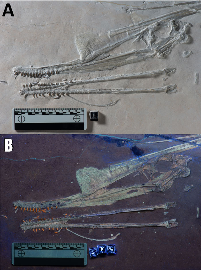
Tracing of the skull of Petrodactyle. Many sutures are uncertain and these labels are for general guidance. Abbreviations are as follows: an, angular; ar, articular; fpc, frontoparietal crest; j, jugal; lc, lacrimal; m, maxilla; n, nasal; naof, nasoantorbital fenestra; o, orbit; p, palate; po, postorbital; ppf, postpalatine fenestra; pm, premaxilla; pmc, premaxillary crest; q, quadrate; qj, quadratojugal; sa, surangular; t, tooth. Scale bar is 100 mm.

Tracing of the skull of Petrodactyle. Many sutures are uncertain and these labels are for general guidance. Abbreviations are as follows: an, angular; ar, articular; fpc, frontoparietal crest; j, jugal; lc, lacrimal; m, maxilla; n, nasal; naof, nasoantorbital fenestra; o, orbit; p, palate; po, postorbital; ppf, postpalatine fenestra; pm, premaxilla; pmc, premaxillary crest; q, quadrate; qj, quadratojugal; sa, surangular; t, tooth. Scale bar is 100 mm.

Many pterosaurs are known with bony crests which they used primarily as sexual signals to other members of the species, but Pterodactyle has by far the largest crest even seen in a ctenochasmatid. Dr David Hone of Queen Mary University of London was the lead author on the study said, “Big though this crest is, we know that these pterosaurs had skin-like extensions attached to it, so in life Petrodactyle would have had an even larger crest.”

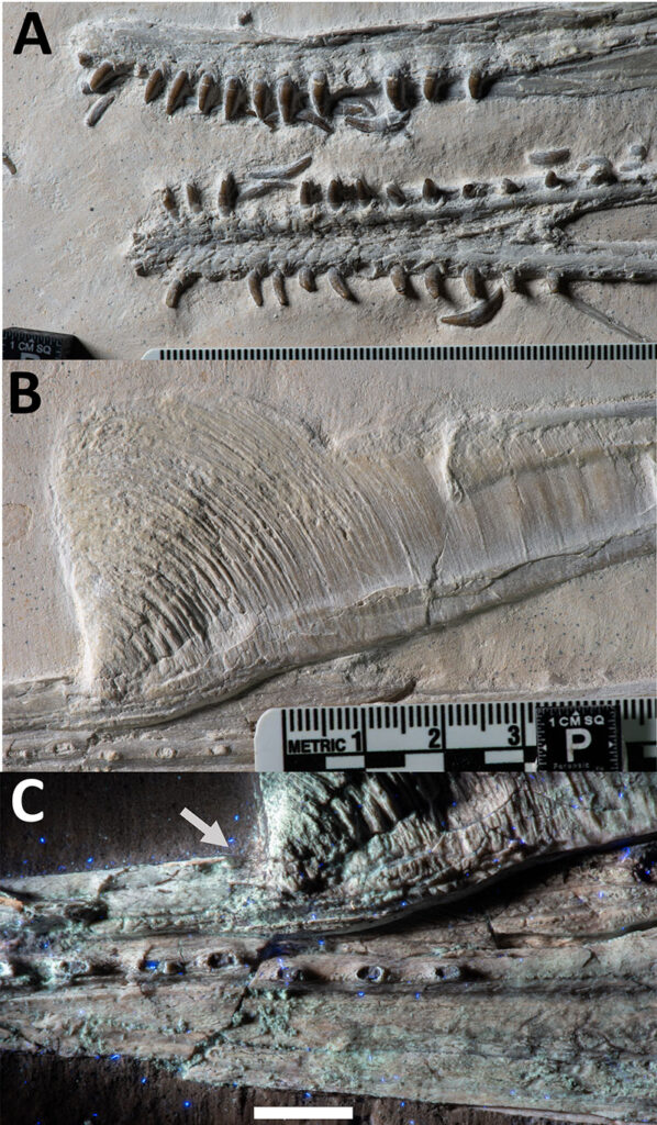
The details of the specimen are especially clear under UV light which helps show the difference between the bones and the rock in which they are embedded, which under natural light are a very similar colour. René Lauer of the Lauer Foundation, an author on the study said, “The use of UV Induced Fluorescence digital photography provided the ability to discern fine structures small bones and provided additional information regarding the structures of the bony crest which aided in the interpretations and conclusions of this unique new species.”

Close up of middle cervical vertebrae of Petrodactyle. A, in left lateral view, and B, in ventral view. Scale bar is 10 mm.

Close up of middle cervical vertebrae of Petrodactyle. A, in left lateral view, and B, in ventral view. Scale bar is 10 mm.

Petrodactyle was unusually large too. It has a wingspan of around 2 meters, but it was still an older ‘teenager’ by pterosaur standards and would have been even larger as a fully mature animal. Even so, it is one of the largest pterosaurs known from the Late Jurassic period. Bruce Lauer of the Lauer Foundation, an author on the study said “The specimen was located in a quarry which is producing scientifically important fossils that provide additional insights into Late Jurassic Pterosaurs. This research is a great example of the benefits of cooperation between amateur collectors, commercial fossil dealers, our Foundation and research scientists to advance science.”

Like other ctenochasmatids, Petrodactyle was at home on the shore of shallow seas but might have ventured into estuaries or to lakes. It’s long jaw with many small teeth would have been good for grabbing at small fish, shrimp and other aquatic prey. However, unlike most other ctenochasmatids, it had an expansion at the back of the skull to attach large jaw muscles and give it a stronger bite than many of its contemporaries. Frederik Spindler of the Dinosaurier Museum in Germany, an author on the study said, “It is amazing to document an increasingly wide range of adaptations. Pterosaurs were a fundamental part of the Jurassic ecology.”

Dr Hone concluded “Peter Wellnhofer is long overdue having a species of German pterosaur named after him to honour his lifelong contribution to the study of these amazing animals.”

The Lauer Foundation acquires, curates, and provides access to a collection of scientifically important Palaeontological specimens. The collection is available to the scientific community for research, publication, exhibition and educational outreach.

  1. David W.E. Hone, René Lauer, Bruce Lauer, Frederik Spindler. Petrodactyle wellnhoferi gen. et sp. nov.: A new and large ctenochasmatid pterosaur from the Late Jurassic of GermanyPalaeontologia Electronica, 2023; DOI: 10.26879/1251
Source: Queen Mary University of London. “New fossil flying reptile ‘Elvis’ takes flight: The 2-meter wingspan animal had a huge bony crest on its head.” ScienceDaily. ScienceDaily, 14 July 2023. <www.sciencedaily.com/releases/2023/07/230714114734.htm>.

@WFS,World Fossil Society, Athira, Riffin T Sajeev,Russel T Sajeev

WFS News: “Achilles Neck” – Fossils Reveal Long-Necked Reptiles Were Decapitated by Predators

@WFS,World Fossil Society, Athira, Riffin T Sajeev,Russel T Sajeev

Artistic rendition of the decapitation scene of Tanystropheus hydroides. Credit: Roc Olivé (Institut Català de Paleontologia Miquel Crusafont)/FECYT

Artistic rendition of the decapitation scene of Tanystropheus hydroides. Credit: Roc Olivé (Institut Català de Paleontologia Miquel Crusafont)/FECYT

Fossil evidence reveals that the long necks of the ancient marine reptiles, Tanystropheus, made them vulnerable to predators. The study found bite marks on the necks of the fossils, providing the first direct proof of this long-suspected evolutionary disadvantage despite their survival success over a span of 175 million years.

In the age of dinosaurs, many marine reptiles had extremely long necks compared to reptiles today. While it was clearly a successful evolutionary strategy, paleontologists have long suspected that their long-necked bodies made them vulnerable to predators. Now, after almost 200 years of continued research, direct fossil evidence confirms this scenario for the first time in the most graphic way imaginable.

Researchers reporting in the journal Current Biology on June 19 studied the unusual necks of two Triassic species of Tanystropheus, a type of reptile distantly related to crocodiles, birds, and dinosaurs. The species had unique necks composed of 13 extremely elongated vertebrae and strut-like ribs. Consequently, these marine reptiles likely possessed stiffened necks and waited to ambush their prey. But Tanystropheus’s predators apparently also took advantage of the long neck for their own gain.

Careful examination of their fossilized bones now shows that the necks of two existing specimens representing different species with severed necks have clear bite marks on them, in one case right where the neck was broken. The findings offer gruesome and exceedingly rare evidence for predator-prey interactions in the fossil record going back over 240 million years ago, the researchers say.

“Paleontologists speculated that these long necks formed an obvious weak spot for predation, as was already vividly depicted almost 200 years ago in a famous painting by Henry de la Beche from 1830,” said Stephan Spiekman of the Staatliches Museum für Naturkunde Stuttgart, Germany. “Nevertheless, there was no evidence of decapitation—or any other sort of attack targeting the neck—known from the abundant fossil record of long-necked marine reptiles until our present study on these two specimens of Tanystropheus.”

Spiekman had studied these reptiles as the main subject of his doctoral work at the Paleontological Museum of the University of Zurich, Switzerland, where the specimens are housed. He recognized that two species of Tanystropheus lived in the same environment, one small species, about a meter and a half in length, likely feeding on soft-shelled animals like shrimp, and a much larger species of up to six meters long that fed on fish and squid. He also found clear evidence in the shape of the skull that Tanystropheus likely spent most of its time in the water.

It had been well known that two specimens of these species had well-preserved heads and necks that abruptly ended. It had been speculated that these necks were bitten off, but no one had studied this in detail. In the new study, Spiekman teamed up with Eudald Mujal, also of the Stuttgart Museum, and a research associate at the Institut Català de Paleontologia Miquel Crusafont, Spain, who is an expert on fossil preservation and predatory interactions in the fossil record based on bite traces on bones. After an afternoon spent examining the two specimens in Zurich, they concluded that the necks had clearly been bitten off.

“Something that caught our attention is that the skull and portion of the neck preserved are undisturbed, only showing some disarticulation due to the typical decay of a carcass in a quiet environment,” Mujal said. “Only the neck and head are preserved; there is no evidence whatsoever of the rest of the animals. The necks end abruptly, indicating they were completely severed by another animal during a particularly violent event, as the presence of tooth traces evinces.”

“The fact that the head and neck are so undisturbed suggests that when they reached the place of their final burial, the bones were still covered by soft tissues like muscle and skin,” Mujal continued. “They were clearly not fed on by the predator. Although this is speculative, it would make sense that the predators were less interested in the skinny neck and small head, and instead focused on the much meatier parts of the body. Taken together, these factors make it most likely that both individuals were decapitated during the hunt and not scavenged, although scavenging can never be fully excluded in fossils that are this old.”

“Interestingly, the same scenario—although certainly executed by different predators—played out for both specimens, which remember, represent individuals of two different Tanystropheus species, which are very different in size and possibly lifestyle,” Spiekman says.

The findings confirm earlier interpretations that the ancient reptiles’ necks represent a completely unique evolutionary structure that was much narrower and stiffer than those of long-necked plesiosaurs, according to the researchers. They also show that evolving a long neck as a sea reptile came with potential downsides. Nevertheless, they note, elongated necks were clearly a highly successful evolutionary strategy, found in many different marine reptiles over a time span of 175 million years.

“In a very broad sense, our research once again shows that evolution is a game of trade-offs,” Spiekman says. “The advantage of having a long neck clearly outweighed the risk of being targeted by a predator for a very long time. Even Tanystropheus itself was quite successful in evolutionary terms, living for at least 10 million years and occurring in what is now Europe, the Middle East, China, North America, and possibly South America.”

Reference: “Decapitation in the long-necked reptile Tanystropheus (Archosauromorpha, Tanystropheidae)” by Stephan N.F. Spiekman and Eudald Mujal, 19 June 2023, Current Biology.
DOI: 10.1016/j.cub.2023.04.027

Source:  Article By Cell Press @ scitechdaily.com

@WFS,World Fossil Society, Athira, Riffin T Sajeev,Russel T Sajeev

WFS News: A new fossil katydid of the genus Arethaea Stål (Orthoptera: Tettigoniidae) with exceptionally preserved internal organs from the Eocene Green River Formation of Colorado

@WFS,World Fossil Society, Athira, Riffin T Sajeev,Russel T Sajeev

A new fossil katydid of the genus Arethaea Stål (Orthoptera: Tettigoniidae) with exceptionally preserved internal organs from the Eocene Green River Formation of Colorado

A new fossil katydid of the genus Arethaea Stål (Orthoptera: Tettigoniidae) with exceptionally preserved internal organs from the Eocene Green River Formation of Colorado

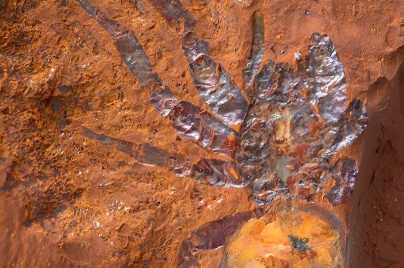
50 million years ago in what is now northwestern Colorado, a katydid died, sank to the bottom of a lake and was quickly buried in fine sediments, where it remained until its compressed fossil was recovered in recent years. When researchers examined the fossil under a microscope, they saw that not only had many of the insect’s hard structures been preserved in the compressed shale, so had several internal organs and tissues, which are not normally fossilized.

They describe their findings in the journal Palaeoentomology.

“Katydids are very rare in the fossil record, so any new katydid fossil you find represents a new data point in the evolutionary history of katydids,” said palaeoentomologist and study lead Sam Heads, the director of the Prairie Research Institute’s Center for Paleontology. “But perhaps the most striking feature of this fossil is the really exceptional, remarkable preservation of internal organs — organs that you just don’t see in fossils.”

The fossil was found in the Green River Formation in Rio Blanco County, Colorado. The formation is vast, extending into three states, and is a famous fossil bed in the western U.S. because the fine-grained shales yield a very detailed record of the plants and animals that once inhabited the region, Heads said.

The katydid belongs to the genus Arethaea, a group today known as thread-legged katydids for their extremely slender, grasslike legs, Heads said. The specimen represents a new but extinct species, which Heads and his colleagues named Arethaea solterae after their colleague Leellen Solter.*

“Obviously, having a fossil species of a modern genus is really significant because it confirms the antiquity of this lineage,” Heads said. “Now we know that about 50 million years ago, this genus had already evolved and already had a morphology that mimics the grass in which it lives and hides from predators.”

The find will help scientists understand how this group of insects evolved and when they developed their unique physical structure, he said.

The rare glimpse of soft internal organs in a 50-million-year-old fossil is also remarkable, Heads said.

“Part of the digestive tract is preserved, a part of the midgut we call the ventriculus,” he said. “That’s not so unusual; we have other specimens from this location that have gut traces, so I wasn’t particularly struck by that.”

But when he looked at the specimen under a microscope, Heads saw evidence of other internal structures that he was not expecting to be preserved. There were traces of the fibers making up thoracic muscles associated with the wings or flank muscles. There was some undifferentiated tissue known as a “fat body,” an organ that aids in insect metabolism.

Even more surprisingly, “there are these little tubules that all seem to connect to a round structure — and that can only be a testis and accessory glands that are associated with the testis,” Heads said. “That’s just phenomenal. I was not expecting to see that kind of structure preserved in a rock compression. I’ve never seen that before.”

To double-check his analysis, Heads dissected several katydid specimens of the same genus to match what he was seeing in the fossil.

“They look exactly the same,” he said. “The testis, the accessory glands and the ventriculus were all the same in the present-day katydids. I was just blown away by it. To my knowledge, this is the first example of this level of preservation.”

  1. Sam W. Heads, M. Jared Thomas, Tyler J. Hedlund, Yinan Wang. A new fossil katydid of the genus Arethaea Stål (Orthoptera: Tettigoniidae) with exceptionally preserved internal organs from the Eocene Green River Formation of ColoradoPalaeoentomology, 2023; 6 (3) DOI: 10.11646/palaeoentomology.6.3.10
 <www.sciencedaily.com/releases/2023/06/230626164301.htm>.

@WFS,World Fossil Society, Athira, Riffin T Sajeev,Russel T Sajeev


WFS News: Megalodon was no cold-blooded killer

@WFS,World Fossil Society, Athira, Riffin T Sajeev,Russel T Sajeev

The largest marine predator that ever lived was no cold-blooded killer.

                   The largest marine predator that ever lived was no cold-blooded killer.

Well, a killer, yes. But a new analysis by environmental scientists from UCLA, UC Merced and William Paterson University sheds light on the warm-blooded animal’s ability to regulate its body temperature — and might help explain why it went extinct.

After analyzing isotopes in the tooth enamel of the ancient shark, which went extinct about 3.6 million years ago, the scientists concluded the megalodon could maintain a body temperature that was about 13 degrees Fahrenheit (about 7 degrees Celsius) warmer than the surrounding water.

That temperature difference is greater than those that have been determined for other sharks that lived alongside the megalodon and is large enough to categorize megalodons as warm-blooded.

The paper, published in Proceedings of the National Academy of Sciences, suggests that the amount of energy the megalodon used to stay warm contributed to its extinction. And it has implications for understanding current and future environmental changes.

“Studying the driving factors behind the extinction of a highly successful predatory shark like megalodon can provide insight into the vulnerability of large marine predators in modern ocean ecosystems experiencing the effects of ongoing climate change,” said lead researcher Robert Eagle, a UCLA assistant professor of atmospheric and oceanic sciences and member of the UCLA Institute of the Environment and Sustainability.

Megalodons, which are believed to have reached lengths up to 50 feet, belonged to a group of sharks called mackerel sharks — members of that group today include the great white and thresher shark. While most fish are cold-blooded, with body temperatures that are the same as the surrounding water, mackerel sharks keep the temperature of all or parts of their bodies somewhat warmer than the water around them, qualities called mesothermy and regional endothermy, respectively.

Sharks store heat generated by their muscles, making them different from fully warm-blooded or endothermic animals like mammals. In mammals, a region of the brain called the hypothalamus regulates body temperature.

Various lines of evidence have hinted that megalodon might have been mesothermic. But without data from the soft tissues that drive body temperature in modern sharks, it has been difficult to determine if or to what extent megalodon was endothermic.

In the new study, the scientists looked for answers in the megalodon’s most abundant fossil remains: its teeth. A main component of teeth is a mineral called apatite, which contains atoms of carbon and oxygen. Like all atoms, carbon and oxygen can come in “light” or “heavy” forms known as isotopes, and the amount of light or heavy isotopes that make up apatite as it forms can depend on a range of environmental factors. So the isotopic composition of fossil teeth can reveal insights about where an animal lived and the types of foods it ate, and — for marine vertebrates — information like the chemistry of the seawater where the animal lived and the animal’s body temperature.

“You can think of the isotopes preserved in the minerals that make up teeth as a kind of thermometer, but one whose reading can be preserved for millions of years,” said Randy Flores, a UCLA doctoral student and fellow of the Center for Diverse Leadership in Science, who worked on the study. “Because teeth form in the tissue of an animal when it’s alive, we can measure the isotopic composition of fossil teeth in order to estimate the temperature at which they formed and that tells us the approximate body temperature of the animal in life.”

Because most ancient and modern sharks are unable to maintain body temperatures significantly higher than the temperature of surrounding seawater, the isotopes in their teeth reflect temperatures that deviate little from the temperature of the ocean. In warm-blooded animals, however, the isotopes in their teeth record the effect of body heat produced by the animal, which is why the teeth indicate temperatures that are warmer than the surrounding seawater.

The researchers hypothesized that any difference between the isotope values of the megalodon and those of other sharks that lived at the same time would indicate the degree to which the megalodon could warm its own body.

The researchers collected teeth from the megalodon and other shark contemporaries from five locations around the world, and analyzed them using mass spectrometers at UCLA and UC Merced. Using statistical modeling to estimate sea water temperatures at each site where teeth were collected, the scientists found that megalodons’ teeth consistently yielded average temperatures that indicated it had an impressive ability to regulate body temperature.

Its warmer body allowed megalodon to move faster, tolerate colder water and spread out around the world. But it was that evolutionary advantage that might have contributed to its downfall, the researchers wrote.

The megalodon lived during the Pliocene Epoch, which began 5.33 million years ago and ended 2.58 million years ago, and global cooling during that period caused sea level and ecological changes that the megalodon did not survive.

“Maintaining an energy level that would allow for megalodon’s elevated body temperature would require a voracious appetite that may not have been sustainable in a time of changing marine ecosystem balances when it may have even had to compete against newcomers such as the great white shark,” Flores said.

Project co-leader Aradhna Tripati, a UCLA professor of Earth, planetary and space sciences and a member of the Institute of Environment and Sustainability, said the scientists now plan to apply the same approach to studying other species.

“Having established endothermy in megalodon, the question arises of how frequently it is found in apex marine predators throughout geologic history,” she said.

  1. Michael L. Griffiths, Robert A. Eagle, Sora L. Kim, Randon J. Flores, Martin A. Becker, Harry M. Maisch, Robin B. Trayler, Rachel L. Chan, Jeremy McCormack, Alliya A. Akhtar, Aradhna K. Tripati, Kenshu Shimada. Endothermic physiology of extinct megatooth sharksProceedings of the National Academy of Sciences, 2023; 120 (27) DOI: 10.1073/pnas.2218153120

Source: <www.sciencedaily.com/releases/2023/06/230626164144.htm>

@WFS,World Fossil Society, Athira, Riffin T Sajeev,Russel T Sajeev